UTI Information Service

Urology health
How your urinary system works.
Your Urinary System is a vital system that helps your body remove waste and excess water through urine. It includes a few key structures that work together seamlessly.
How your urinary system works: An introduction to its key structures
Your urinary system is a vital system that helps your body remove waste and excess water through urine. To understand good urology health it is useful to understand how your urinary system works.
Key structures of your urinary system
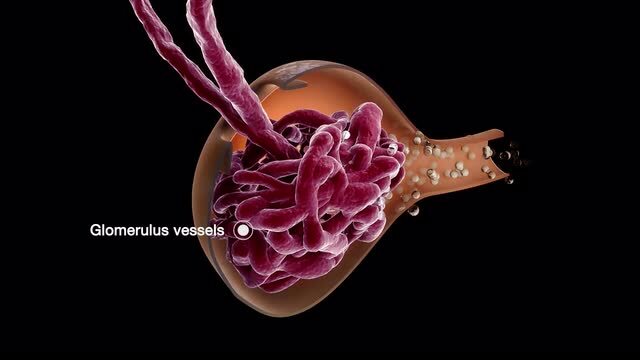
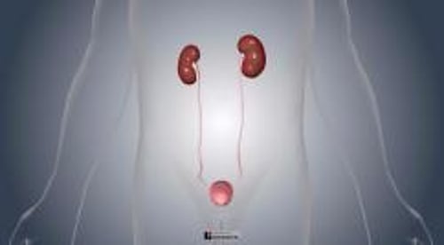
- Kidneys : Located just below your rib cage on either side of your spine, these bean-shaped organs filter blood to remove waste and excess water, creating urine. They also regulate blood pressure and help maintain your body’s balance of minerals.
- Ureters : These narrow tubes transport urine from the kidneys to the bladder. Think of them as the highways for urine travel.
- Bladder : This is like a flexible storage bag that holds urine until you decide it’s time to release it. It expands to store urine and contracts when you urinate.
- Urethra : The final pathway for urine to leave your body. In men, this tube also carries semen and is longer, running through the penis. In women, it’s shorter and opens just above the vaginal opening. It features a valve called the sphincter that prevents leaks until you decide to urinate.
- The urethra is also the point where bacteria can enter the urinary system, potentially leading to infections. Although males do get UTIs, in females the urethra is shorter and closer to the anus, which makes it easier for bacteria from the intestinal tract to reach the bladder.
- Urothelium: Lines most of the urinary tract, including the bladder, some of the urethra, and the ureters, this special lining is remarkably adaptable; it can stretch substantially when your bladder fills with urine and contract when it empties. It acts as a protective barrier and becomes inflamed when exposed to irritants and bacteria. This inflammation can lead to symptoms like pain, urgency, and frequency of urination.
How it all works together
- Filtering : Your kidneys filter your blood, capturing waste and turning it into urine.
- Transporting : Urine travels from your kidneys through the ureters to your bladder.
- Storing : Your bladder holds the urine until you’re ready to go to the bathroom.
- Exiting : When you urinate, the bladder contracts, and urine exits through the urethra.
The health of your urinary system is crucial for maintaining your body’s natural balance and efficiency. A urinary tract infection (UTI), which most commonly affects the bladder and urethra, can disrupt these processes.
UTIs and your urinary system
Understanding what UTIs are, why they happen, and how they affect you is vital for early recognition, effective treatment, and taking steps to try to prevent them. This knowledge is essential, whether you’re looking after your own health or supporting someone else’s.
Your Urinary System includes a few key structures that work together seamlessly. Understanding these structures can help you grasp how UTIs can develop and affect your body.
For more information about UTIs, click here .
Key facts about UTIs
- 50-60 % of females in the UK will experience at least 1 UTI in their lifetime and those who have more than three in a year are more likely to continue to have them.
- Males experience UTI’s too and are usually associated with obstruction in the urinary tract, usually the prostate.
- Individual risk factors can increase the likelihood of UTIs.
- UTIs can range from simple and occasional to complex and recurrent.
Urinary Tract Infections (UTI)
Urinary Tract Infections (UTIs) are a major health concern affecting millions globally each year. These infections not only cause discomfort but can also significantly impact daily life and overall well-being. See our information on UTIs
UTI Information Hub and Helpline
Welcome to Our UTI Information Hub and Helpline. These resources are dedicated to empowering, supporting, and informing those affected by UTIs, including patients, their relatives, and the general public
Your urology health relates to the 'urogenital system' in both males and females. These are the parts of your body responsible for producing, storing and discharging urine (kidneys, bladder, urethra) and the parts of your body involved in sexual function (prostate, penis, and testicles)
Find out more
Get involved.
Whether it's funding research, supporting urology healthcare professionals, raising awareness of urology health or campaigning, we couldn't do it without your support
Impact & highlights
In the past eight years alone we have invested over £5.5 million in high-calibre research projects, which have made significant contributions to the understanding of urology cancers and conditions
Sign up to our newsletter
An official website of the United States government
Here’s how you know
Official websites use .gov A .gov website belongs to an official government organization in the United States.
Secure .gov websites use HTTPS A lock ( Lock Locked padlock icon ) or https:// means you’ve safely connected to the .gov website. Share sensitive information only on official, secure websites.
- Entire Site
- Research & Funding
- Health Information
- About NIDDK
- Urologic Diseases
- The Urinary Tract & How It Works
- Español
The Urinary Tract & How It Works
On this page:
What is the urinary tract?
How does urination occur, why is the urinary tract important, what affects the amount of urine you produce, how can you keep your urinary tract healthy, clinical trials.
The urinary tract is the body’s drainage system for removing urine , which is made up of wastes and extra fluid. For normal urination to occur, all body parts in the urinary tract need to work together, and in the correct order.
The urinary tract includes two kidneys , two ureters , a bladder , and a urethra .
Kidneys . Two bean-shaped organs, each about the size of a fist. They are located just below your rib cage, one on each side of your spine. Every day, your kidneys filter about 120 to 150 quarts of blood to remove wastes and balance fluids. This process produces about 1 to 2 quarts of urine per day.
Ureters . Thin tubes of muscle that connect your kidneys to your bladder and carry urine to the bladder.
Bladder . A hollow, muscular, balloon-shaped organ that expands as it fills with urine. The bladder sits in your pelvis between your hip bones. A normal bladder acts like a reservoir. It can hold 1.5 to 2 cups of urine. Although you do not control how your kidneys function, you can control when to empty your bladder. Bladder emptying is known as urination.
Urethra . A tube located at the bottom of the bladder that allows urine to exit the body during urination.

The urinary tract includes two sets of muscles that work together as a sphincter , closing off the urethra to keep urine in the bladder between your trips to the bathroom.
- The internal sphincter muscles of the bladder neck and urethra stay closed until your brain sends signals to urinate.
- The external sphincter muscles surround the internal sphincter and provide extra pressure to keep the urethra closed. You can consciously squeeze the external sphincter and the pelvic floor muscles to keep urine from leaking out.
To urinate, your brain signals the sphincters to relax. Then it signals the muscular bladder wall to tighten, squeezing urine through the urethra and out of your bladder.
How often you need to urinate depends on how quickly your kidneys produce the urine that fills the bladder and how much urine your bladder can comfortably hold. The muscles of your bladder wall remain relaxed while the bladder fills with urine, and the sphincter muscles remain contracted to keep urine in the bladder. As your bladder fills up, signals sent to your brain tell you to find a toilet soon.
The urinary tract is important because it filters wastes and extra fluid from the bloodstream and removes them from the body.
The amount of urine you produce depends on many factors, such as the amount of liquid and food you consume and the amount of fluid you lose through sweating and breathing. Certain medicines, medical conditions, and types of food can also affect the amount of urine you produce. Children produce less urine than adults.
You can help keep your urinary tract healthy by following some basic tips.
Drink enough liquids, especially water . If you’re healthy, try to drink six to eight 8-ounce glasses of liquid each day. You may need to drink more if you have kidney stones or bladder stones. At least half of your liquid intake should be water. You might need to drink less water if you have certain conditions, such as kidney failure or heart disease . Ask your health care professional how much liquid is healthy for you.
Keep your bowels regular . Regular bowel movements are important to your bladder health. You can promote both bowel health and bladder health by
- making healthy food choices . You can keep your urinary tract healthy by sticking to an eating plan that includes lean proteins, whole grains, fiber -rich breads, nuts, colorful berries, fruits, and vegetables to promote regular bowel movements.
- living a healthy lifestyle . Get regular physical activity , limit your alcohol intake, cut down on caffeinated food and drinks, and don’t smoke.
Go whenever you need to . Often, people will hold their urine because it’s not a good time to go to the bathroom. However, holding in your urine for too long can weaken your bladder muscles and make it harder for your bladder to empty completely. Urine left in your bladder can allow bacteria to grow and makes you more likely to develop a urinary tract infection (UTI) .
Develop healthy bathroom habits . Take enough time to fully empty your bladder when urinating—don’t rush it. Urinate after sex to flush away bacteria that may have entered the urethra during sex. Clean the genital area before and after sex. If you’re a woman, wipe from front to back, especially after a bowel movement, to keep bacteria from getting into the urethra.
Stay in tune with your body . Pay attention to how often you feel the urge to urinate. Take note if you need to urinate more often than usual, if your urine leaks, if it becomes more difficult for you to begin urinating, or if you feel you’re not able to completely empty your bladder. These changes may be early signs of different urinary tract problems. Talk with your health care professional if you notice any of these signs. You may be able to prevent a condition from becoming more severe if you get help early on.
Do pelvic floor muscle exercises . Pelvic floor exercises, also called Kegel exercises , can keep your pelvic floor muscles strong and maintain healthy bladder and bowel function. Both men and women can benefit from pelvic floor muscle exercises.

The NIDDK conducts and supports clinical trials in many diseases and conditions, including urologic diseases. The trials look to find new ways to prevent, detect, or treat disease and improve quality of life.
What are clinical trials, and are they right for you?
Watch a video of NIDDK Director Dr. Griffin P. Rodgers explaining the importance of participating in clinical trials.
This content is provided as a service of the National Institute of Diabetes and Digestive and Kidney Diseases (NIDDK), part of the National Institutes of Health. NIDDK translates and disseminates research findings to increase knowledge and understanding about health and disease among patients, health professionals, and the public. Content produced by NIDDK is carefully reviewed by NIDDK scientists and other experts.
The NIDDK would like to thank: Ariana L. Smith, M.D., FPMRS, University of Pennsylvania Health System
25.8 Urine Transport and Elimination
Key takeaways.
By the end of this section, you will be able to:
Describe how the kidney modifies filtrate to influence urine production
- Describe the characteristics of a normal urine sample
- Explain the role of the loop of Henle, the vasa recta, and the countercurrent multiplication mechanisms in urine production
- Explain the role of aldosterone and ADH in urine production
- Identify the ureters, urinary bladder, and urethra, as well as their location, structure, histology, and function
- Describe the micturition reflex
- Describe voluntary and involuntary neural control of micturition
Urine is the end product once the filtrate has been fully manipulated by the nephrons. Until the filtrate passes through the renal papilla into the minor calyx, it can be affected by nephron processes. This is how kidneys produce anywhere from .4 L of urine/day to as much as 20L urine/day, all while balancing plasma composition and excreting potential toxins in the urine.
Composition of Urine
The two kidneys filter your entire blood volume many times each day to remove wastes as urine. Characteristics of urine can be variable ( Table 25.1 ) depending on water intake and losses, nutrient intake, and other factors described in this chapter, though cells, proteins and blood are not normally found in the urine. Some of the characteristics such as color and odor are rough descriptors of your state of hydration. For example, if you exercise or work outside, and sweat a great deal, your urine will turn darker and produce a slight odor. Alternatively, a well hydrated person will have light or clear colored urine with little odor ( Figure 25.8.1 ).

The pH (hydrogen ion concentration) of the urine can vary more than 1000-fold, from a normal low of 4.5 to a maximum of 8.0 depending on actions of specific cells of the kidney. Urine osmolarity is the number of osmoles or milliosmoles per liter of fluid (mOsmol/L). Urine osmolarity ranges from a low of 50–100 mOsmol/L to as high as 1200 mOsmol/L H 2 O. The color of urine is determined mostly by the breakdown products of red blood cell destruction ( Figure 25.8.1 ). The “heme” of hemoglobin is converted by the liver into water-soluble forms that can be excreted into the bile and indirectly into the urine. This yellow pigment is urochrome .
Urine color may also be affected by certain foods like beets, berries, and fava beans. Dehydration produces darker, concentrated urine that may also possess the slight odor of ammonia. Ammonia (NH 3 ) is a toxic byproduct of protein metabolism. It is formed as amino acids are deaminated by liver hepatocytes. That means that the amine group, NH 2 , is removed from amino acids as they are broken down. Most of the resulting ammonia is converted into urea by liver hepatocytes. Urea is not only less toxic but is utilized to aid in the recovery of water by the loop of Henle and collecting ducts to control urine volume.
Urine volume varies considerably. The normal range is one to two liters per day. The kidneys must produce a minimum urine volume of about 400 mL/day to rid the body of wastes. Output below this level may be caused by severe dehydration or renal disease. The regulation of urine volume reflects regulation of urine and blood composition as described below.
Blood is filtered, and the filtrate is transformed into urine at a relatively constant rate throughout the day. Urine is stored until a convenient time for excretion. All structures involved in the transport and storage of the urine are large enough to be visible to the naked eye. This transport and storage system not only stores the waste, but it protects the tissues from damage due to the wide range of pH and osmolarity of the urine.
As urine is formed, it drains into the calyces of the kidney, which merge to form the funnel-shaped renal pelvis in the hilum of each kidney. The renal pelvis narrows to become the ureter of each kidney. As urine passes through the ureter, it does not passively drain into the bladder but rather is propelled by waves of peristalsis. As the ureters enter the pelvis, they pass laterally, hugging the pelvic walls. As they approach the bladder, they turn medially and join with the bladder wall obliquely. This is important because it creates an one-way valve (a physiological sphincter rather than an anatomical sphincter ) that allows urine into the bladder but prevents reflux of urine from the bladder back into the ureter. Children born lacking this oblique course of the ureter through the bladder wall are susceptible to “vesicoureteral reflux,” which dramatically increases their risk of serious UTI. Pregnancy also increases the likelihood of reflux and UTI.
The ureters are approximately 30 cm long. The inner mucosa is lined with transitional epithelium ( Figure 25.8.2 ) and scattered goblet cells that secrete protective mucus. The muscular layer of the ureter consists of longitudinal and circular smooth muscles that create the peristaltic contractions to move the urine into the bladder without the aid of gravity. Finally, a loose adventitial layer composed of collagen and fat anchors the ureters between the parietal peritoneum and the posterior abdominal wall.

The urinary bladder collects urine from both ureters ( Figure 25.8.3 ). The bladder lies posterior to the pubic bone and anterior to the rectum. The bladder is partially retroperitoneal (outside the peritoneal cavity) with its peritoneal-covered “dome” projecting into the abdomen when the bladder is distended with urine. When empty, the region of the bladder that does not collapse is called the trigone (Greek tri- = “triangle” and the root of the word “trigonometry”), which is delineated by the opening of the ureters and the urethra, forming a triangular area.

External Website

View the University of Michigan WebScope at http://141.214.65.171/Histology/Urinary%20System/212N_HISTO_40X.svs/view.apml to explore the tissue sample in greater detail.
The bladder is a highly distensible organ comprised of irregular crisscrossing bands of smooth muscle collectively called the detrusor muscle . The interior surface is made of transitional cellular epithelium that is structurally suited for the large volume fluctuations of the bladder. When empty, it resembles columnar epithelia, but when stretched, it “transitions” (hence the name) to a squamous appearance (see Figure 25.8.3 ). Volumes in adults can range from nearly zero to 500–600 mL.
The detrusor muscle contracts with significant force in the young. The bladder’s strength diminishes with age, but voluntary contractions of abdominal skeletal muscles can increase intra-abdominal pressure to promote more forceful bladder emptying. Such voluntary contraction is also used in forceful defecation and childbirth.
The urethra transports urine from the bladder to the outside of the body for disposal. The urethra is the only urologic organ that shows any significant anatomic difference between males and females; all other urine transport structures are identical ( Figure 25.8.4 ).

The urethra in both males and females begins inferior and central to the trigone. The urethra tracks posterior and inferior to the pubic symphysis (see Figure 25.8.4 a ). In both males and females, the proximal urethra is lined by transitional epithelium, whereas the terminal portion is a nonkeratinized, stratified squamous epithelium. In the male, pseudostratified columnar epithelium lines the urethra between these two cell types. Voiding is regulated by an involuntary autonomic nervous system-controlled internal urinary sphincter , consisting of smooth muscle and voluntary skeletal muscle that forms the external urinary sphincter below it. In females, the urethra’s short length, about 4 cm, is less of a barrier to fecal bacteria than the longer male urethra and the best explanation for the greater incidence of UTI in women. Voluntary control of the external urethral sphincter is a function of the pudendal nerve. It arises in the sacral region of the spinal cord, traveling via the S2–S4 nerves of the sacral plexus.
Micturition Reflex
Micturition is the physiological term for urination or voiding. It results from an interplay of involuntary and voluntary actions by the internal and external urethral sphincters. When bladder volume reaches about 150 mL, an urge to void is sensed but is easily overridden. Voluntary control of urination relies on consciously preventing relaxation of the external urethral sphincter to maintain urinary continence. As the bladder fills, subsequent urges become harder to ignore.
Micturition is a result of stretch receptors in the bladder wall that transmit nerve impulses to the sacral region of the spinal cord to generate a spinal reflex. The resulting parasympathetic neural outflow causes contraction of the detrusor muscle and relaxation of the involuntary internal urethral sphincter. At the same time, the spinal cord inhibits somatic motor neurons, resulting in the relaxation of the skeletal muscle of the external urethral sphincter. The micturition reflex is active in infants but with maturity, children learn to override the reflex by asserting external sphincter control, thereby delaying voiding (potty training).
Nerves involved in the control of urination include the hypogastric, pelvic, and pudendal ( Figure 25.8.5 ). Voluntary micturition requires an intact spinal cord and functional pudendal nerve arising from the sacral micturition center . Since the external urinary sphincter is voluntary skeletal muscle, actions by cholinergic neurons maintain contraction (and thereby continence) during filling of the bladder. At the same time, sympathetic nervous activity via the hypogastric nerves suppresses contraction of the detrusor muscle. With further bladder stretch, afferent signals traveling over sacral pelvic nerves activate parasympathetic neurons. This activates efferent neurons to release acetylcholine at the neuromuscular junctions, producing detrusor contraction and bladder emptying.

Chapter Review
The urethra is the only urinary structure that differs significantly between males and females. This is due to the dual role of the male urethra in transporting both urine and semen. The urethra arises from the trigone area at the base of the bladder. Urination is controlled by an involuntary internal sphincter of smooth muscle and a voluntary external sphincter of skeletal muscle. The shorter female urethra contributes to the higher incidence of bladder infections in females. The bladder is largely retroperitoneal and can hold up to 500–600 mL urine. Micturition is the process of voiding the urine and involves both involuntary and voluntary actions. Voluntary control of micturition requires a mature and intact sacral micturition center. It also requires an intact spinal cord. Loss of control of micturition is called incontinence and results in voiding when the bladder contains about 250 mL urine. The ureters are retroperitoneal and lead from the renal pelvis of the kidney to the trigone area at the base of the bladder. A thick muscular wall consisting of longitudinal and circular smooth muscle helps move urine toward the bladder by way of peristaltic contractions.
Review Questions
1. Peristaltic contractions occur in the ________.
- urethra, bladder, and ureters
2. Somatic motor neurons must be ________ to relax the external urethral sphincter to allow urination.
3. Which part of the urinary system is not completely retroperitoneal?
Critical Thinking Questions
1. Why are females more likely to contract bladder infections than males?
2. Describe how forceful urination is accomplished.
Answers for Review Questions
Answers for Critical Thinking Questions
- The longer urethra of males means bacteria must travel farther to the bladder to cause an infection.
- Forceful urination is accomplished by contraction of abdominal muscles.
This work, Anatomy & Physiology, is adapted from Anatomy & Physiology by OpenStax , licensed under CC BY . This edition, with revised content and artwork, is licensed under CC BY-SA except where otherwise noted.
Images, from Anatomy & Physiology by OpenStax , are licensed under CC BY except where otherwise noted.
Access the original for free at https://openstax.org/books/anatomy-and-physiology/pages/1-introduction .
Anatomy & Physiology Copyright © 2019 by Lindsay M. Biga, Staci Bronson, Sierra Dawson, Amy Harwell, Robin Hopkins, Joel Kaufmann, Mike LeMaster, Philip Matern, Katie Morrison-Graham, Kristen Oja, Devon Quick, Jon Runyeon, OSU OERU, and OpenStax is licensed under a Creative Commons Attribution-ShareAlike 4.0 International License , except where otherwise noted.

- school Campus Bookshelves
- menu_book Bookshelves
- perm_media Learning Objects
- login Login
- how_to_reg Request Instructor Account
- hub Instructor Commons
Margin Size
- Download Page (PDF)
- Download Full Book (PDF)
- Periodic Table
- Physics Constants
- Scientific Calculator
- Reference & Cite
- Tools expand_more
- Readability
selected template will load here
This action is not available.
24.5A: Overview of Urine Transport, Storage, and Elimination
- Last updated
- Save as PDF
- Page ID 8151
\( \newcommand{\vecs}[1]{\overset { \scriptstyle \rightharpoonup} {\mathbf{#1}} } \)
\( \newcommand{\vecd}[1]{\overset{-\!-\!\rightharpoonup}{\vphantom{a}\smash {#1}}} \)
\( \newcommand{\id}{\mathrm{id}}\) \( \newcommand{\Span}{\mathrm{span}}\)
( \newcommand{\kernel}{\mathrm{null}\,}\) \( \newcommand{\range}{\mathrm{range}\,}\)
\( \newcommand{\RealPart}{\mathrm{Re}}\) \( \newcommand{\ImaginaryPart}{\mathrm{Im}}\)
\( \newcommand{\Argument}{\mathrm{Arg}}\) \( \newcommand{\norm}[1]{\| #1 \|}\)
\( \newcommand{\inner}[2]{\langle #1, #2 \rangle}\)
\( \newcommand{\Span}{\mathrm{span}}\)
\( \newcommand{\id}{\mathrm{id}}\)
\( \newcommand{\kernel}{\mathrm{null}\,}\)
\( \newcommand{\range}{\mathrm{range}\,}\)
\( \newcommand{\RealPart}{\mathrm{Re}}\)
\( \newcommand{\ImaginaryPart}{\mathrm{Im}}\)
\( \newcommand{\Argument}{\mathrm{Arg}}\)
\( \newcommand{\norm}[1]{\| #1 \|}\)
\( \newcommand{\Span}{\mathrm{span}}\) \( \newcommand{\AA}{\unicode[.8,0]{x212B}}\)
\( \newcommand{\vectorA}[1]{\vec{#1}} % arrow\)
\( \newcommand{\vectorAt}[1]{\vec{\text{#1}}} % arrow\)
\( \newcommand{\vectorB}[1]{\overset { \scriptstyle \rightharpoonup} {\mathbf{#1}} } \)
\( \newcommand{\vectorC}[1]{\textbf{#1}} \)
\( \newcommand{\vectorD}[1]{\overrightarrow{#1}} \)
\( \newcommand{\vectorDt}[1]{\overrightarrow{\text{#1}}} \)
\( \newcommand{\vectE}[1]{\overset{-\!-\!\rightharpoonup}{\vphantom{a}\smash{\mathbf {#1}}}} \)
The urinary organs include the kidneys, ureters, bladder, and urethra.
Learning Objectives
- Outline the process of urine transport, storage, and elimination
- Urine collects from the nephrons and flows into the ureters.
- The ureters use smooth muscle contractions to facilitate the flow of urine.
- The urinary bladder is a hollow, muscular, and elastic organ that stores urine.
- Urine exits the bladder and the body through the urethra.
- The kidneys, ureters, bladder, and urethra make up the urinary tract, the pathway through which urine flows and is eliminated from the body.
- ureter : These are two long, narrow ducts that carry urine from the kidneys to the urinary bladder.
- urinary bladder : An elastic, muscular sac situated in the pelvic cavity, into which urine from the kidneys is stored prior to disposal by urination. Urine enters the bladder via the ureters and exits via the urethra.
The Urinary System

Urinary tract : The transport and removal of urine from the body follows the urinary tract.
The organs, tubes, muscles, and nerves that work together to create, store, and carry urine are referred to as the urinary system, which is another name for the renal system. The renal system filters the plasma of blood and regulates blood volume by excreting excess water in the form of urine. Urine transport follows a path through the kidneys, ureters, bladder, and urethra, which are collectively known as the urinary tract.
Urine Transport
Urine is essentially water, ions, and secreted molecules that leave the collecting duct of the many nephrons of the kidney and flow into the ureters. The ureters are two tubes that drain urine from the kidneys to the bladder.
Each ureter is a muscular tube that drains into the bladder. Smooth muscle contractions in the walls of the ureters, over time, send the urine in small spurts into the bladder, the organ where urine is stored before it can be eliminated.
Urine Storage
The bladder is a hollow muscular organ shaped like a balloon. It sits in the pelvis and is held in place by ligaments attached to other organs and the pelvic bones. The bladder stores urine until enough of it accumulates for removal from the body. It swells into a round shape when it is full and gets smaller when empty.
If the urinary system is healthy, the bladder can hold up to 16 ounces (2 cups) of urine comfortably for 2 to 5 hours. Circular muscles called sphincters help keep urine from leaking. The sphincter muscles close tightly, like a rubber band, around the opening of the bladder into the urethra, the tube that allows urine to pass outside the body.
Urine Elimination
Nerves in the bladder are stimulated as the bladder fills with urine and becomes larger, which in turn stimulates the need to urinate. When you urinate, the brain signals the bladder muscles to tighten, squeezing urine out of the bladder.
At the same time, the brain signals the sphincter muscles to relax. As these muscles relax, urine exits the bladder through the urethra, and leaves the body through an opening in the genital region that contains the urethra. When all the signals occur in the correct order, normal urination occurs, removing urine from the body.

- Search the Collections or Website

- Search the Collections or Website -->
- Urology A-Z
The Urinary Tract System
Attention: Restrictions on use of AUA, AUAER, and UCF content in third party applications, including artificial intelligence technologies, such as large language models and generative AI. You are prohibited from using or uploading content you accessed through this website into external applications, bots, software, or websites, including those using artificial intelligence technologies and infrastructure, including deep learning, machine learning and large language models and generative AI.
What is Urology?
Urology is a part of health care that deals with a lot of different body parts. This includes body parts that form the Urinary System and Male Reproductive System. If you have a problem with a body part in these two systems, you may need to see a urologist.
The Urinary System
Many of your body parts work with each other to form the Urinary System. Urine is taken out of the body if these parts work with each other in the right order. This allows normal urination to happen. For both men and women, the main parts of the system are Kidneys, Ureters, Bladder and Urethra. Urine is produced in the kidneys. It flows through tubes called ureters, and into the bladder. Urine leaves the body through the urethra.
How the Kidneys Work
The kidneys are fist-size organs that make urine. They are found on both sides of the spine behind the liver, stomach, pancreas and bowels. Healthy kidneys work like clockwork to turn extra water and waste into urine.
How the Ureters Work
Urine flows out of the kidneys and into the ureters. Ureters are thin tubes of muscle that connect the kidneys to the bladder. Ureters carry urine from the kidneys to the bladder.
How the Bladder Works
The bladder is a hollow, balloon-shaped organ. It is mostly made of muscle. It stores urine until you are ready to go to the bathroom to release it. The bladder helps you urinate. The brain tells it to tighten and force the urine out.
How the Urethra Works
Urine leaves the body through a hollow tube connected to the bladder. This tube is called a urethra.
The Male Reproductive System
Many body parts work with each other to form the Male Reproductive System. The purpose is for each part to work in the right order so a male can have sex. During sex, you may be able to fertilize a woman's ovum (egg) and make a baby. Not all men are able to have sex, even if their Male Reproductive System works right.
How the Testicles Work
The testicles (also known as testes) are two golf ball size glands held in a sac (scrotum) below the penis. The testicles have a firm, slightly spongy feel. At the top and outside edge is a rubbery, tube-like structure called the epididymis. The firmness of the teste should be the same throughout. The size of the testicles should be about the same.
The testicles make male hormones. The most common hormone is testosterone, which controls the sex drive (libido). It also triggers the development of male traits, such as facial hair. The testicles also make sperm, the male reproductive cells, which travel through a group of tube-like structures to the epididymis. Sperm cells are then carried from the testicles by the vas deferens to the seminal vesicles, where they are mixed with fluid from the prostate gland.
How the Prostate Works
The prostate is a walnut-shaped gland inside the male body. The prostate sits under the bladder and in front of the rectum. The prostate's main job is to help make fluid for semen to help protect and energize the sperm as they travel to the female egg.
During ejaculation the sperm cells, seminal vesicle fluid and prostate fluid enter the urethra (the tube in the penis through which urine and seminal fluid leave the body).
How the Penis Works
The penis carries sperm out of the body. There are three tubes inside the penis. One is called the urethra. It is hollow and carries urine from the bladder through the penis to the outside. The other two tubes are called the corpora cavernosa. These are spongy tubes that are soft until filled with blood during an erection. The three tubes are wrapped together by a very tough fibrous sheath called the tunica albuginea.
During sex, the stiffness of the penis makes it hard enough to be inserted into the woman's vagina. In this case, the urethra acts as a tube for semen to be ejaculated into the vagina. When you ejaculate, seminal fluid and seminal vesicles mix with sperm to form semen. The semen travels through the urethra and comes out the end of your penis.
Explore Further

We're On a Global Mission!

Why a Clinical Trial Might Be Right for You
This website uses cookies. We use cookies to enable you to more easily use our website, to monitor and analyze the use of our site to help improve our website and services, and to assist us with advertising reporting functions. By checking the “I Agree” box, you consent to our use of cookies as described in our Privacy Policy. I Agree You can learn more about our Cookie Policy here
Your Browser is Not Supported
This web site has been optimized for user experience and security, therefore Internet Explorer(IE) is not a recommended browser.Please use the latest version of Microsoft Edge, Chrome, Firefox or Safari(MacOS). Thank you.
25.2 Gross Anatomy of Urine Transport
Learning objectives.
By the end of this section, you will be able to:
- Identify the ureters, urinary bladder, and urethra, as well as their location, structure, histology, and function
- Compare and contrast male and female urethras
- Describe the micturition reflex
- Describe voluntary and involuntary neural control of micturition
Rather than start with urine formation, this section will start with urine excretion. Urine is a fluid of variable composition that requires specialized structures to remove it from the body safely and efficiently. Blood is filtered, and the filtrate is transformed into urine at a relatively constant rate throughout the day. This processed liquid is stored until a convenient time for excretion. All structures involved in the transport and storage of the urine are large enough to be visible to the naked eye. This transport and storage system not only stores the waste, but it protects the tissues from damage due to the wide range of pH and osmolarity of the urine, prevents infection by foreign organisms, and for the male, provides reproductive functions.
The urethra transports urine from the bladder to the outside of the body for disposal. The urethra is the only urologic organ that shows any significant anatomic difference between males and females; all other urine transport structures are identical ( Figure 25.3 ).
The urethra in both males and females begins inferior and central to the two ureteral openings forming the three points of a triangular-shaped area at the base of the bladder called the trigone (Greek tri- = “triangle” and the root of the word “trigonometry”). The urethra tracks posterior and inferior to the pubic symphysis (see Figure 25.3 a ). In both males and females, the proximal urethra is lined by transitional epithelium, whereas the terminal portion is a nonkeratinized, stratified squamous epithelium. In the male, pseudostratified columnar epithelium lines the urethra between these two cell types. Voiding is regulated by an involuntary autonomic nervous system-controlled internal urinary sphincter , consisting of smooth muscle and voluntary skeletal muscle that forms the external urinary sphincter below it.
Female Urethra
The external urethral orifice is embedded in the anterior vaginal wall inferior to the clitoris, superior to the vaginal opening (introitus), and medial to the labia minora. Its short length, about 4 cm, is less of a barrier to fecal bacteria than the longer male urethra and the best explanation for the greater incidence of UTI in women. Voluntary control of the external urethral sphincter is a function of the pudendal nerve. It arises in the sacral region of the spinal cord, traveling via the S2–S4 nerves of the sacral plexus.
Male Urethra
The male urethra passes through the prostate gland immediately inferior to the bladder before passing below the pubic symphysis (see Figure 25.3 b ). The length of the male urethra varies between men but averages 20 cm in length. It is divided into four regions: the preprostatic urethra, the prostatic urethra, the membranous urethra, and the spongy or penile urethra. The preprostatic urethra is very short and incorporated into the bladder wall. The prostatic urethra passes through the prostate gland. During sexual intercourse, it receives sperm via the ejaculatory ducts and secretions from the seminal vesicles. Paired Cowper’s glands (bulbourethral glands) produce and secrete mucus into the urethra to buffer urethral pH during sexual stimulation. The mucus neutralizes the usually acidic environment and lubricates the urethra, decreasing the resistance to ejaculation. The membranous urethra passes through the deep muscles of the perineum, where it is invested by the overlying urethral sphincters. The spongy urethra exits at the tip (external urethral orifice) of the penis after passing through the corpus spongiosum. Mucous glands are found along much of the length of the urethra and protect the urethra from extremes of urine pH. Innervation is the same in both males and females.
The urinary bladder collects urine from both ureters ( Figure 25.4 ). The bladder lies anterior to the uterus in females, posterior to the pubic bone and anterior to the rectum. During late pregnancy, its capacity is reduced due to compression by the enlarging uterus, resulting in increased frequency of urination. In males, the anatomy is similar, minus the uterus, and with the addition of the prostate inferior to the bladder. The bladder is a retroperitoneal organ whose "dome" distends superiorly when the bladder is filling with urine.
Interactive Link
View the University of Michigan WebScope to explore the tissue sample in greater detail.
The bladder is a highly distensible organ comprised of irregular crisscrossing bands of smooth muscle collectively called the detrusor muscle . The interior surface is made of transitional cellular epithelium that is structurally suited for the large volume fluctuations of the bladder. When empty, it resembles columnar epithelia, but when stretched, it “transitions” (hence the name) to a squamous appearance (see Figure 25.4 ). Volumes in adults can range from nearly zero to 500–600 mL.
The detrusor muscle contracts with significant force in the young. The bladder’s strength diminishes with age, but voluntary contractions of abdominal skeletal muscles can increase intra-abdominal pressure to promote more forceful bladder emptying. Such voluntary contraction is also used in forceful defecation and childbirth.
Micturition Reflex
Micturition is a less-often used, but proper term for urination or voiding. It results from an interplay of involuntary and voluntary actions by the internal and external urethral sphincters. When bladder volume reaches about 150 mL, an urge to void is sensed but is easily overridden. Voluntary control of urination relies on consciously preventing relaxation of the external urethral sphincter to maintain urinary continence. As the bladder fills, subsequent urges become harder to ignore. Ultimately, voluntary constraint fails with resulting incontinence , which will occur as bladder volume approaches 300 to 400 mL.
Normal micturition is a result of stretch receptors in the bladder wall that transmit nerve impulses to the sacral region of the spinal cord to generate a spinal reflex. The resulting parasympathetic neural outflow causes contraction of the detrusor muscle and relaxation of the involuntary internal urethral sphincter. At the same time, the spinal cord inhibits somatic motor neurons, resulting in the relaxation of the skeletal muscle of the external urethral sphincter. The micturition reflex is active in infants but with maturity, children learn to override the reflex by asserting external sphincter control, thereby delaying voiding (potty training). This reflex may be preserved even in the face of spinal cord injury that results in paraplegia or quadriplegia. However, relaxation of the external sphincter may not be possible in all cases, and therefore, periodic catheterization may be necessary for bladder emptying.
Nerves involved in the control of urination include the hypogastric, pelvic, and pudendal ( Figure 25.5 ). Voluntary micturition requires an intact spinal cord and functional pudendal nerve arising from the sacral micturition center . Since the external urinary sphincter is voluntary skeletal muscle, actions by cholinergic neurons maintain contraction (and thereby continence) during filling of the bladder. At the same time, sympathetic nervous activity via the hypogastric nerves suppresses contraction of the detrusor muscle. With further bladder stretch, afferent signals traveling over sacral pelvic nerves activate parasympathetic neurons. This activates efferent neurons to release acetylcholine at the neuromuscular junctions, producing detrusor contraction and bladder emptying.
The kidneys and ureters are completely retroperitoneal, and the bladder has a peritoneal covering only over the dome. As urine is formed, it drains into the calyces of the kidney, which merge to form the funnel-shaped renal pelvis in the hilum of each kidney. The renal pelvis narrows to become the ureter of each kidney. As urine passes through the ureter, it does not passively drain into the bladder but rather is propelled by waves of peristalsis. As the ureters enter the pelvis, they sweep laterally, hugging the pelvic walls. As they approach the bladder, they turn medially and pierce the bladder wall obliquely. This is important because it creates a one-way valve (a physiological sphincter rather than an anatomical sphincter ) that allows urine into the bladder but prevents reflux of urine from the bladder back into the ureter. Children born lacking this oblique course of the ureter through the bladder wall are susceptible to “vesicoureteral reflux,” which dramatically increases their risk of serious UTI. Pregnancy also increases the likelihood of reflux and UTI.
The ureters are approximately 30 cm long. The inner mucosa is lined with transitional epithelium ( Figure 25.6 ) and scattered goblet cells that secrete protective mucus. The muscular layer of the ureter consists of longitudinal and circular smooth muscles that create the peristaltic contractions to move the urine into the bladder without the aid of gravity. Finally, a loose adventitial layer composed of collagen and fat anchors the ureters between the parietal peritoneum and the posterior abdominal wall.
As an Amazon Associate we earn from qualifying purchases.
This book may not be used in the training of large language models or otherwise be ingested into large language models or generative AI offerings without OpenStax's permission.
Want to cite, share, or modify this book? This book uses the Creative Commons Attribution License and you must attribute OpenStax.
Access for free at https://openstax.org/books/anatomy-and-physiology/pages/1-introduction
- Authors: J. Gordon Betts, Kelly A. Young, James A. Wise, Eddie Johnson, Brandon Poe, Dean H. Kruse, Oksana Korol, Jody E. Johnson, Mark Womble, Peter DeSaix
- Publisher/website: OpenStax
- Book title: Anatomy and Physiology
- Publication date: Apr 25, 2013
- Location: Houston, Texas
- Book URL: https://openstax.org/books/anatomy-and-physiology/pages/1-introduction
- Section URL: https://openstax.org/books/anatomy-and-physiology/pages/25-2-gross-anatomy-of-urine-transport
© Jan 27, 2022 OpenStax. Textbook content produced by OpenStax is licensed under a Creative Commons Attribution License . The OpenStax name, OpenStax logo, OpenStax book covers, OpenStax CNX name, and OpenStax CNX logo are not subject to the Creative Commons license and may not be reproduced without the prior and express written consent of Rice University.
Chapter 9: The Urinary System
Ureters, urinary bladder, and urethra.
Urine is a fluid of variable composition that requires specialized structures to remove it from the body safely and efficiently. Blood is filtered, and the filtrate is transformed into urine at a relatively constant rate throughout the day. This processed liquid is stored until a convenient time for excretion. All structures involved in the transport and storage of the urine are large enough to be visible to the naked eye. This transport and storage system not only stores the waste, but it protects the tissues from damage due to the wide range of pH and osmolarity of the urine, prevents infection by foreign organisms, and for the male, provides reproductive functions.
The kidneys and ureters are completely retroperitoneal, and the bladder has a peritoneal covering only over the dome. As urine is formed, it drains into the calyces of the kidney, which merge to form the funnel-shaped renal pelvis in the hilum of each kidney. The hilum narrows to become the ureter of each kidney. As urine passes through the ureter, it does not passively drain into the bladder but rather is propelled by waves of peristalsis. As the ureters enter the pelvis, they sweep laterally, hugging the pelvic walls. As they approach the bladder, they turn medially and pierce the bladder wall obliquely. This is important because it creates a one-way valve (a physiological sphincter rather than an anatomical sphincter ) that allows urine into the bladder but prevents reflux of urine from the bladder back into the ureter. Children born lacking this oblique course of the ureter through the bladder wall are susceptible to “vesicoureteral reflux,” which dramatically increases their risk of serious UTI. Pregnancy also increases the likelihood of reflux and UTI.
The ureters are approximately 30 cm long. The inner mucosa is lined with transitional epithelium (Figure 1) and scattered goblet cells that secrete protective mucus. The muscular layer of the ureter consists of longitudinal and circular smooth muscles that create the peristaltic contractions to move the urine into the bladder without the aid of gravity. Finally, a loose adventitial layer composed of collagen and fat anchors the ureters between the parietal peritoneum and the posterior abdominal wall.

Figure 1. Peristaltic contractions help to move urine through the lumen with contributions from fluid pressure and gravity. LM × 128. (Micrograph provided by the Regents of the University of Michigan Medical School © 2012)
Urinary Bladder
The urinary bladder collects urine from both ureters. The bladder lies anterior to the uterus in females, posterior to the pubic bone and anterior to the rectum. During late pregnancy, its capacity is reduced due to compression by the enlarging uterus, resulting in increased frequency of urination. In males, the anatomy is similar, minus the uterus, and with the addition of the prostate inferior to the bladder. The bladder is partially retroperitoneal (outside the peritoneal cavity) with its peritoneal-covered “dome” projecting into the abdomen when the bladder is distended with urine.

Figure 2. (a) Anterior cross section of the bladder. (b) The detrusor muscle of the bladder (source: monkey tissue) LM × 448. (Micrograph provided by the Regents of the University of Michigan Medical School © 2012)
The bladder is a highly distensible organ comprised of irregular crisscrossing bands of smooth muscle collectively called the detrusor muscle . The interior surface is made of transitional cellular epithelium that is structurally suited for the large volume fluctuations of the bladder. When empty, it resembles columnar epithelia, but when stretched, it “transitions” (hence the name) to a squamous appearance. Volumes in adults can range from nearly zero to 500–600 mL.
The detrusor muscle contracts with significant force in the young. The bladder’s strength diminishes with age, but voluntary contractions of abdominal skeletal muscles can increase intra-abdominal pressure to promote more forceful bladder emptying. Such voluntary contraction is also used in forceful defecation and childbirth.
Micturition Reflex
Micturition is a less-often used, but proper term for urination or voiding. It results from an interplay of involuntary and voluntary actions by the internal and external urethral sphincters. When bladder volume reaches about 150 mL, an urge to void is sensed but is easily overridden. Voluntary control of urination relies on consciously preventing relaxation of the external urethral sphincter to maintain urinary continence. As the bladder fills, subsequent urges become harder to ignore. Ultimately, voluntary constraint fails with resulting incontinence , which will occur as bladder volume approaches 300 to 400 mL.
Normal micturition is a result of stretch receptors in the bladder wall that transmit nerve impulses to the sacral region of the spinal cord to generate a spinal reflex. The resulting parasympathetic neural outflow causes contraction of the detrusor muscle and relaxation of the involuntary internal urethral sphincter. At the same time, the spinal cord inhibits somatic motor neurons, resulting in the relaxation of the skeletal muscle of the external urethral sphincter. The micturition reflex is active in infants but with maturity, children learn to override the reflex by asserting external sphincter control, thereby delaying voiding (potty training). This reflex may be preserved even in the face of spinal cord injury that results in paraplegia or quadriplegia. However, relaxation of the external sphincter may not be possible in all cases, and therefore, periodic catheterization may be necessary for bladder emptying.
Nerves involved in the control of urination include the hypogastric, pelvic, and pudendal. Voluntary micturition requires an intact spinal cord and functional pudendal nerve arising from the sacral micturition center . Since the external urinary sphincter is voluntary skeletal muscle, actions by cholinergic neurons maintain contraction (and thereby continence) during filling of the bladder. At the same time, sympathetic nervous activity via the hypogastric nerves suppresses contraction of the detrusor muscle. With further bladder stretch, afferent signals traveling over sacral pelvic nerves activate parasympathetic neurons. This activates efferent neurons to release acetylcholine at the neuromuscular junctions, producing detrusor contraction and bladder emptying.

Figure 3. Nerves Innervating the Urinary System
The urethra transports urine from the bladder to the outside of the body for disposal. The urethra is the only urologic organ that shows any significant anatomic difference between males and females; all other urine transport structures are identical.

Figure 4. The urethra transports urine from the bladder to the outside of the body. This image shows (a) a female urethra and (b) a male urethra.
The urethra in both males and females begins inferior and central to the two ureteral openings forming the three points of a triangular-shaped area at the base of the bladder called the trigone (Greek tri – = “triangle” and the root of the word “trigonometry”). The urethra tracks posterior and inferior to the pubic symphysis (see Figure 4). In both males and females, the proximal urethra is lined by transitional epithelium, whereas the terminal portion is a nonkeratinized, stratified squamous epithelium. In the male, pseudostratified columnar epithelium lines the urethra between these two cell types. Voiding is regulated by an involuntary autonomic nervous system-controlled internal urinary sphincter , consisting of smooth muscle and voluntary skeletal muscle that forms the external urinary sphincter below it.
Female Urethra
The external urethral orifice is embedded in the anterior vaginal wall inferior to the clitoris, superior to the vaginal opening (introitus), and medial to the labia minora. Its short length, about 4 cm, is less of a barrier to fecal bacteria than the longer male urethra and the best explanation for the greater incidence of UTI in women. Voluntary control of the external urethral sphincter is a function of the pudendal nerve. It arises in the sacral region of the spinal cord, traveling via the S2–S4 nerves of the sacral plexus.
Male Urethra
The male urethra passes through the prostate gland immediately inferior to the bladder before passing below the pubic symphysis (see Figure 4b). The length of the male urethra varies between men but averages 20 cm in length. It is divided into three regions: the prostatic urethra, the membranous urethra, and the spongy or penile urethra. The prostatic urethra passes through the prostate gland. During sexual intercourse, it receives sperm via the ejaculatory ducts and secretions from the seminal vesicles. Paired Cowper’s glands (bulbourethral glands) produce and secrete mucus into the urethra to buffer urethral pH during sexual stimulation. The mucus neutralizes the usually acidic environment and lubricates the urethra, decreasing the resistance to ejaculation. The membranous urethra passes through the deep muscles of the perineum, where it is invested by the overlying urethral sphincters. The spongy urethra exits at the tip (external urethral orifice) of the penis after passing through the corpus spongiosum. Mucous glands are found along much of the length of the urethra and protect the urethra from extremes of urine pH. Innervation is the same in both males and females.
Chapter Review
The urethra is the only urinary structure that differs significantly between males and females. This is due to the dual role of the male urethra in transporting both urine and semen. The urethra arises from the trigone area at the base of the bladder. Urination is controlled by an involuntary internal sphincter of smooth muscle and a voluntary external sphincter of skeletal muscle. The shorter female urethra contributes to the higher incidence of bladder infections in females. The male urethra receives secretions from the prostate gland, Cowper’s gland, and seminal vesicles as well as sperm. The bladder is largely retroperitoneal and can hold up to 500–600 mL urine. Micturition is the process of voiding the urine and involves both involuntary and voluntary actions. Voluntary control of micturition requires a mature and intact sacral micturition center. It also requires an intact spinal cord. Loss of control of micturition is called incontinence and results in voiding when the bladder contains about 250 mL urine. The ureters are retroperitoneal and lead from the renal pelvis of the kidney to the trigone area at the base of the bladder. A thick muscular wall consisting of longitudinal and circular smooth muscle helps move urine toward the bladder by way of peristaltic contractions.
anatomical sphincter: smooth or skeletal muscle surrounding the lumen of a vessel or hollow organ that can restrict flow when contracted
detrusor muscle: smooth muscle in the bladder wall; fibers run in all directions to reduce the size of the organ when emptying it of urine
external urinary sphincter: skeletal muscle; must be relaxed consciously to void urine
internal urinary sphincter: smooth muscle at the juncture of the bladder and urethra; relaxes as the bladder fills to allow urine into the urethra
incontinence: loss of ability to control micturition
micturition: also called urination or voiding
physiological sphincter: sphincter consisting of circular smooth muscle indistinguishable from adjacent muscle but possessing differential innervations, permitting its function as a sphincter; structurally weak
retroperitoneal: outside the peritoneal cavity; in the case of the kidney and ureters, between the parietal peritoneum and the abdominal wall
sacral micturition center: group of neurons in the sacral region of the spinal cord that controls urination; acts reflexively unless its action is modified by higher brain centers to allow voluntary urination
trigone: area at the base of the bladder marked by the two ureters in the posterior–lateral aspect and the urethral orifice in the anterior aspect oriented like points on a triangle
urethra: transports urine from the bladder to the outside environment
- View All Systems
- Anatomy Systems
- Cardiovascular
- Respiratory
- Integumentary
- View All Telehealth
- Online Pharmacies
- Hims Review
- Keeps Review
- Keeps vs Hims
- BlueChew Review
- Rugiet Review
- Hims ED Review
- Online Therapy
- BetterHelp Review
- BetterHelp vs Talkspace
- View All Testing
- General Health Tests
- Best DNA Health Test
- Best Microbiome Test
- Viome Review
- Best At-Home Herpes Test
- STDcheck Review
- View All Self-Care
- Hair and Hair Loss
- Best Hair Loss Treatment for Men
- Best Laser Cap for Hair Loss
- Kiierr Laser Cap Review
- Nutrition Guide
- Found Weight Loss Review
- Relationships
- Best STD Dating Websites
- Best Herpes Dating Websites
- Sexual Health
- Best Erectile Dysfunction Treatment
- Best Delay Spray
- Best Prostate Massager
- BlueChew Free Trial
- Best CBD for Arthritis
- Best Incontinence Underwear for Women
- Best Incontinence Underwear for Men
- All Health Products
- Best Amino Acid Supplements
- Best Muscle Building Stack
- Best NMN Supplement
- Best NAC Supplement
- Best NAD Supplement
- Best Berberine Supplement
- Mood and Energy
- Best Nootropics
- Best Saffron Supplement
- Fitness and Weight
- Vital Reds Review
- Total Restore Review
- Best Testosterone Booster
- Best Male Enhancement Pills
- Best Volume Pills
- View All Research
- Skeletal System
- Muscular System
- Cardiovascular System
- Digestive System
- Nervous System
- Respiratory System
- Urinary System
The Urinary System
Explore the anatomy and physiology of the kidneys, ureters, urinary bladder, and urethra, learning all about how our bodies filter out and remove waste through urine..
- 2D Interactive
- NEW 3D Rotate and Zoom
Anatomy Explorer
- LOWER TORSO
Urinary Bladder
Change current view angle.
- Urinary System (Male View)
- Urinary System (Male Posterior View)
- Urinary System (Posterior View)
Toggle Anatomy System
- Endocrine System
- Female Reproductive System
- Immune and Lymphatic Systems
- Integumentary System
- Male Reproductive System
Switch Gender
Anatomy term.
Displayed on other page

The urinary system consists of the kidneys, ureters, urinary bladder, and urethra. The kidneys filter the blood to remove wastes and produce urine. The ureters, urinary bladder, and urethra together form the urinary tract, which acts as a plumbing system to drain urine from the kidneys, store it, and then release it during urination. Besides filtering and eliminating wastes from the body, the urinary system also maintains the homeostasis of water, ions, pH, blood pressure, calciummycontentbreak and red blood cells.
Urinary System Anatomy
The kidneys are a pair of bean-shaped organs found along the posterior wall of the abdominal cavity. The left kidney is located slightly higher than the right kidney because the right side of the liver is much larger than the left side. The kidneys, unlike the other organs of the abdominal cavity, are located posterior to the peritoneum and touch the muscles of the back . The kidneys are surrounded by a layer of adipose that holds them in place and protects them from physical damage. The kidneys filter metabolic wastes, excess ions, and chemicals from the blood to form urine.
The ureters are a pair of tubes that carry urine from the kidneys to the urinary bladder. The ureters are about 10 to 12 inches long and run on the left and right sides of the body parallel to the vertebral column . Gravity and peristalsis of smooth muscle tissue in the walls of the ureters move urine toward the urinary bladder. The ends of the ureters extend slightly into the urinary bladder and are sealed at the point of entry to the bladder by the ureterovesical valves. These valves prevent urine from flowing back towards the kidneys.
The urinary bladder is a sac-like hollow organ used for the storage of urine. The urinary bladder is located along the body’s midline at the inferior end of the pelvis . Urine entering the urinary bladder from the ureters slowly fills the hollow space of the bladder and stretches its elastic walls. The walls of the bladder allow it to stretch to hold anywhere from 600 to 800 milliliters of urine.
The urethra is the tube through which urine passes from the bladder to the exterior of the body. The female urethra is around 2 inches long and ends inferior to the clitoris and superior to the vaginal opening. In males, the urethra is around 8 to 10 inches long and ends at the tip of the penis . The urethra is also an organ of the male reproductive system as it carries sperm out of the body through the penis.

The flow of urine through the urethra is controlled by the internal and external urethral sphincter muscles. The internal urethral sphincter is made of smooth muscle and opens involuntarily when the bladder reaches a certain set level of distention. The opening of the internal sphincter results in the sensation of needing to urinate. The external urethral sphincter is made of skeletal muscle and may be opened to allow urine to pass through the urethra or may be held closed to delay urination.
Urinary System Physiology
Maintenance of homeostasis.
The kidneys maintain the homeostasis of several important internal conditions by controlling the excretion of substances out of the body.
The kidney can control the excretion of potassium, sodium, calcium, magnesium, phosphate, and chloride ions into urine. In cases where these ions reach a higher than normal concentration, the kidneys can increase their excretion out of the body to return them to a normal level. Conversely, the kidneys can conserve these ions when they are present in lower than normal levels by allowing the ions to be reabsorbed into the blood during filtration. (See more about ions .)
The kidneys monitor and regulate the levels of hydrogen ions (H+) and bicarbonate ions in the blood to control blood pH. H+ ions are produced as a natural byproduct of the metabolism of dietary proteins and accumulate in the blood over time. The kidneys excrete excess H+ ions into urine for elimination from the body. The kidneys also conserve bicarbonate ions, which act as important pH buffers in the blood.
The cells of the body need to grow in an isotonic environment in order to maintain their fluid and electrolyte balance. The kidneys maintain the body’s osmotic balance by controlling the amount of water that is filtered out of the blood and excreted into urine. When a person consumes a large amount of water, the kidneys reduce their reabsorption of water to allow the excess water to be excreted in urine. This results in the production of dilute, watery urine. In the case of the body being dehydrated, the kidneys reabsorb as much water as possible back into the blood to produce highly concentrated urine full of excreted ions and wastes. The changes in excretion of water are controlled by antidiuretic hormone (ADH). ADH is produced in the hypothalamus and released by the posterior pituitary gland to help the body retain water.
Blood Pressure
The kidneys monitor the body’s blood pressure to help maintain homeostasis. When blood pressure is elevated, the kidneys can help to reduce blood pressure by reducing the volume of blood in the body. The kidneys are able to reduce blood volume by reducing the reabsorption of water into the blood and producing watery, dilute urine. When blood pressure becomes too low, the kidneys can produce the enzyme renin to constrict blood vessels and produce concentrated urine, which allows more water to remain in the blood.
Inside each kidney are around a million tiny structures called nephrons. The nephron is the functional unit of the kidney that filters blood to produce urine. Arterioles in the kidneys deliver blood to a bundle of capillaries surrounded by a capsule called a glomerulus . As blood flows through the glomerulus, much of the blood’s plasma is pushed out of the capillaries and into the capsule, leaving the blood cells and a small amount of plasma to continue flowing through the capillaries. The liquid filtrate in the capsule flows through a series of tubules lined with filtering cells and surrounded by capillaries. The cells surrounding the tubules selectively absorb water and substances from the filtrate in the tubule and return it to the blood in the capillaries. At the same time, waste products present in the blood are secreted into the filtrate. By the end of this process, the filtrate in the tubule has become urine containing only water, waste products, and excess ions. The blood exiting the capillaries has reabsorbed all of the nutrients along with most of the water and ions that the body needs to function.
Storage and Excretion of Wastes
After urine has been produced by the kidneys, it is transported through the ureters to the urinary bladder. The urinary bladder fills with urine and stores it until the body is ready for its excretion. When the volume of the urinary bladder reaches anywhere from 150 to 400 milliliters, its walls begin to stretch and stretch receptors in its walls send signals to the brain and spinal cord . These signals result in the relaxation of the involuntary internal urethral sphincter and the sensation of needing to urinate. Urination may be delayed as long as the bladder does not exceed its maximum volume, but increasing nerve signals lead to greater discomfort and desire to urinate.
Urination is the process of releasing urine from the urinary bladder through the urethra and out of the body. The process of urination begins when the muscles of the urethral sphincters relax, allowing urine to pass through the urethra. At the same time that the sphincters relax, the smooth muscle in the walls of the urinary bladder contract to expel urine from the bladder.
Production of Hormones
The kidneys produce and interact with several hormones that are involved in the control of systems outside of the urinary system.
Calcitriol is the active form of vitamin D in the human body. It is produced by the kidneys from precursor molecules produced by UV radiation striking the skin. Calcitriol works together with parathyroid hormone (PTH) to raise the level of calcium ions in the bloodstream. When the level of calcium ions in the blood drops below a threshold level, the parathyroid glands release PTH, which in turn stimulates the kidneys to release calcitriol. Calcitriol promotes the small intestine to absorb calcium from food and deposit it into the bloodstream. It also stimulates the osteoclasts of the skeletal system to break down bone matrix to release calcium ions into the blood.
Erythropoietin
Erythropoietin, also known as EPO, is a hormone that is produced by the kidneys to stimulate the production of red blood cells. The kidneys monitor the condition of the blood that passes through their capillaries, including the oxygen-carrying capacity of the blood. When the blood becomes hypoxic, meaning that it is carrying deficient levels of oxygen, cells lining the capillaries begin producing EPO and release it into the bloodstream. EPO travels through the blood to the r ed bone marrow , where it stimulates hematopoietic cells to increase their rate of red blood cell production. Red blood cells contain hemoglobin, which greatly increases the blood’s oxygen-carrying capacity and effectively ends the hypoxic conditions.
Renin is not a hormone itself, but an enzyme that the kidneys produce to start the renin-angiotensin system (RAS). The RAS increases blood volume and blood pressure in response to low blood pressure, blood loss, or dehydration. Renin is released into the blood where it catalyzes angiotensinogen from the liver into angiotensin I. Angiotensin I is further catalyzed by another enzyme into Angiotensin II.
Angiotensin II stimulates several processes, including stimulating the adrenal cortex to produce the hormone aldosterone. Aldosterone then changes the function of the kidneys to increase the reabsorption of water and sodium ions into the blood, increasing blood volume and raising blood pressure. Negative feedback from increased blood pressure finally turns off the RAS to maintain healthy blood pressure levels.
Other Recommended Resources
- BetterHelp Reviews
- Fast HSDD Treatments for Women
- Quick Extender Pro Reviews
- Best Extender
- Best Vacuum Pump for Erectile Dysfunction
- STDcheck Reviews
- Herpes Dating Sites
- Types of Bones
- Axial Skeleton
- Appendicular Skeleton
- Skeletal Pathologies
- Muscle Tissue Types
- Muscle Attachments & Actions
- Muscle Contractions
- Muscular Pathologies
- Functions of Blood
- Blood Vessels
- Circulation
- Circulatory Pathologies
- Respiratory Functions
- Upper Respiratory System
- Lower Respiratory System
- Respiratory Pathologies
- Oral Cavity
- Alimentary Canal
- Accessory Organs
- Absorption & Elimination
- Digestive Pathologies
- Lymphatic Structures
- Immune System
Urinary System Structures
- Urine Formation
- Urine Storage & Elimination
- Urinary Pathologies
- Female Reproductive System
- Male Reproductive System
- Reproductive Process
- Endocrine Glands
Urine Storage and Elimination: The Path of Urine Through the Body
© 2024 Visible Body
Urinary bladder

Urine produced in the kidneys travels down the ureters into the urinary bladder. The bladder expands like an elastic sac to hold more urine. As it reaches capacity, the process of micturition, or urination, begins. Involuntary muscle movements send signals to the nervous system, putting the decision to urinate under conscious control.
1. The Two Urethral Sphincters Maintain Urinary Continence

The internal urethral sphincter and the external urethral sphincter both provide muscle control for the flow of urine. The internal sphincter is involuntary. It surrounds the opening of the bladder to the urethra and relaxes to allow urine to pass. The external sphincter is voluntary. It surrounds the urethra outside the bladder and must be relaxed for urination to occur.
2. The Bladder Expands As It Fills with Urine

The bladder is shaped like a pyramid when empty. It becomes more oval as it fills with urine and expands. A smooth muscle called the detrusor surrounds the bladder, and folds called rugae line the interior wall. These structures give the bladder elasticity and allow it to expand. The floor of the bladder includes a funnel-like region called the trigone , formed by the two ureteral orifices and the internal urethral sphincter. Urine flows into the bladder via the ureteral orifices and out through the internal sphincter.
3. Muscles of Micturition: The Detrusor and Urethral Sphincters

Micturition, or urination, is the act of emptying the bladder. When the bladder is full of urine, stretch receptors in the bladder wall trigger the micturition reflex. The detrusor muscle that surrounds the bladder contracts. The internal urethral sphincter relaxes, allowing for urine to pass out of the bladder into the urethra. Both of these reactions are involuntary. The external urethral sphincter is voluntary. It must be relaxed for urine to flow through the urethra and outside the body.
4. How Full Does the Bladder Get Before We Have to Urinate?

The bladder expands as urine flows in from the ureters, but there is a limit to the volume it can contain. At about 200 ml of urine, the detrusor muscle begins to contract and the internal urethral sphincter muscle begins to relax. This sends signals through the nervous system and creates the “urge” to urinate. If this urge is ignored, continence may be threatened. At about 500 ml, detrusor muscle contractions begin to force open the internal urethral sphincter. Unless the external urethral sphincter is powerful enough to prevent it, micturition (urination) will occur involuntarily.
5. Bladder Stretch Triggers the Nervous System to Initiate and Control Micturition

Smooth muscle stretch initiates the micturition reflex by activating stretch receptors in the bladder wall. This autonomic reflex causes the detrusor muscle to contract and the internal urethral sphincter muscle to relax, allowing urine to flow into the urethra. The stretch receptors also send a message to the thalamus and the cerebral cortex, giving voluntary control over the external urethral sphincter. We usually gain this control of urination between the ages of 2 and 3, as our brains develop.
Download Urinary System Lab Manual
External Sources
A description of the urinary bladder from the 1918 edition of Gray's Anatomy of the Human Body.
Using bladder ultrasound to detect urinary retention in patients from Nursing Times .
Visible Body Web Suite provides in-depth coverage of each body system in a guided, visually stunning presentation.
Related Articles
Give It Up for the Kidneys
The Three Steps of Urine Formation
Common Diseases and Disorders

For students
For instructors
Get our awesome anatomy emails!
When you select "Subscribe" you will start receiving our email newsletter. Use the links at the bottom of any email to manage the type of emails you receive or to unsubscribe. See our privacy policy for additional details.
- User Agreement
- Permissions
Overview of the Urinary Tract
Normally, a person has two kidneys . The rest of the urinary tract consists of the following:
Two ureters (the tubes connecting each kidney to the bladder)
The bladder (an expandable muscular sac that holds urine until it is released from the body)
The urethra (a tube attached to the bladder that leads to the outside of the body)
Each kidney continuously produces urine, which then drains through the ureter into the bladder at a low pressure. From the bladder, urine drains through the urethra and exits the body through the penis in males and at the vulva (the area of the external female genital organs) in females. Usually, urine is free of bacteria and other infectious organisms.
Organs of the Urinary Tract
- Cookie Preferences

Copyright © 2024 Merck & Co., Inc., Rahway, NJ, USA and its affiliates. All rights reserved.

An official website of the United States government
The .gov means it's official. Federal government websites often end in .gov or .mil. Before sharing sensitive information, make sure you're on a federal government site.
The site is secure. The https:// ensures that you are connecting to the official website and that any information you provide is encrypted and transmitted securely.
- Publications
- Account settings
- Browse Titles
NCBI Bookshelf. A service of the National Library of Medicine, National Institutes of Health.
StatPearls [Internet]. Treasure Island (FL): StatPearls Publishing; 2024 Jan-.

StatPearls [Internet].
Anatomy, abdomen and pelvis, male genitourinary tract.
Eric H. Wu ; Franco L. De Cicco .
Affiliations
Last Update: September 12, 2022 .
- Introduction
The male genitourinary tract is responsible for the excretion of urine, produced by the kidney, and semen from the sperm from the testes and the various accessory glands. The urethra serves as the final path of excretion of both urine and semen from the body. See Image. Male Genitourinary Tract.
Urine is the waste product of filtered blood that leaves the kidney via the collecting system. From the renal collecting system, urine is transported through the ureters and stored in the urinary bladder for elimination through the urethra.
Sperm is produced in the testes and undergoes maturation in the epididymis. During ejaculation, sperm moves through the epididymis to the vas deferens. The vas deferens is the duct for transporting sperm from the epididymis. It is in the scrotal sac and passes through the inguinal canal into the abdominopelvic cavity. As the vas deferens course to the posterior portion of the prostate, they join the seminal vesicles. The seminal vesicle ducts add seminal fluid to the ejaculate. The area of convergence of the seminal vesicles and vas deferens forms the ejaculatory ducts.
The ejaculatory ducts enter the prostate and empty into the prostatic urethra. Within the prostatic urethra, additional secretions from the prostate gland itself are added to the seminal fluid. The final addition of ejaculatory fluid is a lubricating mucus from the bulbourethral glands, located in the membranous urethra. At ejaculation, the semen leaves through the urethra.
- Structure and Function
The genitourinary tract begins at the kidney, a retroperitoneal organ located at T12-L3 whose primary function is to filter blood and secrete waste products as urine. Urine, produced in the glomeruli and modified in the tubules, eventually travels to the kidney's hilum, emptying into the ureters. The ureters are 25 to 30-cm-long muscular ducts that transport urine to the bladder via peristaltic contractions. They travel down the pelvis, crossing anteriorly to the common iliac artery. When the ureters reach the ischial spine level, they curve anteromedially to enter the bladder at an oblique angle. This angle prevents the backflow of urine by collapsing the ureters when the bladder fills.
The urinary bladder is a hollow, muscular organ composed of the detrusor muscle lined with a specialized distensible epithelium urothelium (previously called the transitional epithelium). The interior of the empty bladder displays an undulating surface, except for a triangular-shaped region, the trigone, defined by the entrance of the left and right ureters and the internal urethral orifice. The trigone has stretch receptors for the sensation of bladder fullness. The bladder is situated posterior to the pubic symphysis in the pelvic cavity. Between the bladder and the pubic bone is a potential space filled with fat called the pre-vesicle space or space of Retzius.
Sperm begins meiosis in the seminiferous tubules and travels to the efferent ductules through the rete testis, anastomosing network located in the mediastinum testes. The efferent ductules connect the rete testis to the epididymis, where the sperm matures. During ejaculation, mature sperm travel through the vas deferens, which ascend through the inguinal canal into the pelvic cavity, looping posterolaterally to the prostate.
The urethra comprises 4 segments named based on its location relative to other organs. The pre-prostatic portion of the urethra is between the prostate and the bladder. The prostatic urethra runs through the prostate. The membranous urethra is located in the deep perineal pouch, along with the bulbourethral gland and external urethral sphincter. Finally, the spongy urethra, surrounded by the corpus spongiosum muscle of the penis, is the last segment reached before expelling the fluid via the urinary meatus. The prostate, a walnut-sized structure located between the bladder and penis, secretes an alkaline solution containing nutrients to mix with the incoming sperm and seminal fluid.
The internal urethral orifice has a smooth muscle sphincter, the internal urethral sphincter, that tonically contracts via the action of the sympathetic nervous system to control continence. [1] Passage through the membranous urethra, located in the deep perineal pouch, is regulated by the external urethral sphincter, which controls the voluntary opening of the membranous urethra by the pudendal nerve from S2-S4. [2] In addition to preventing the outflow of urine from the bladder, the internal urethra sphincter also serves to prevent the backflow of incoming semen from the prostatic urethra into the bladder.
The urinary and genital tract originates initially from the intermediate mesoderm, which differentiates into the urogenital ridge and further differentiates into the nephrogenic and gonadal cords, respectively. The nephrogenic cord develops into the pronephros, mesonephros, and metanephros. Most of the pronephros degenerate. The mesonephric duct forms the ureteric bud, which branches to form the major tracts of the kidneys, including the collecting ducts and the ureter. The metanephros forms the bulk of the kidney, such as the glomerulus. [3] As the kidney develops, it ascends from S1-S2 to T12-L3 while rotating 90 degrees so that the hilum, which initially faces ventrally, faces medially. The cloaca is an endodermal lined structure that divides into the urogenital sinus, which forms the bladder while the trigone forms from Wolffian ducts. [4]
Until fourth week of the development, the gonad is undifferentiated. Male or female differentiation does not start until the seventh week of gestation. [5] The presence of the Y chromosome, specifically of an area called the testes determining factor (SRY) signals to cause the formation of the testes from the undifferentiated gonads. The Leydig cells within the developing testes secrete testosterone to support the maintenance of the male embryological ducts, the mesonephric ducts (Wolffian ducts). [6]
Additionally, the androgens produced by the Leydig cells are responsible for the development of the male external genitals. Also, within the male gonad are Sertoli cells, which secrete anti-Mullerian hormone to inhibit the development of the paramesonephric ducts (Mullerian ducts), the pathway for female development. The mesonephric duct forms the epididymis, vas deferens, and seminal glands. [7]
The mesonephric ducts also give rise to the ureteric bud and trigone of the bladder in both sexes. The ureters and collecting system of the kidney develop from the ureteric bud. The prostate and urethra are both formed from the urogenital sinus. The primordial phallus forms from the genital tubercle in the cranial end of the cloacal membrane. At around 25 to 26 weeks of development, the testicles descend transabdominally, through the inguinal canal, into the scrotum guided by the gubernaculum. The gubernaculum is the genito-inguinal ligament that develops in the 7th week of mesenchymal origin. [8]
- Blood Supply and Lymphatics
The proximal portion of the genitourinary tract, consisting of both kidneys and upper ureters, is supplied by the renal arteries, which are branches of the abdominal aorta, located at the L1-L4 vertebrate. The majority of the distal genitourinary tract, including the distal ureters, bladder, and urethra, receives vascular supply via the superior and inferior vesical arteries, which are branches of the internal iliac artery. [9] The superior and inferior vesical arteries also supply the vas deferens and anastomoses with the testicular artery.
The testes and epididymis arterial supply, venous, and lymphatic drainage reflect the path of descent of the gonad. As the testes descend through the spermatic cord, the relevant artery, veins, and lymphatics follow the testes into the scrotum. The lymphatics of the pelvic organs derive their names from their associated blood vessels. The 4 primary pelvic lymph nodes are located adjacent to the pelvis, consisting of the external iliac, internal iliac, sacral, and common iliac lymph nodes. [10] The external portion of the genitals drains to the superficial inguinal nodes while the internal genital organs drain to the lumbar and paraaortic nodes.
Parasympathetic stimulation on the urinary tract causes relaxation of internal sphincters of the bladder while contracting the detrusor muscle, allowing urination. Conversely, sympathetic stimulation of the urinary tract contracts the internal sphincters of the bladder while relaxing the detrusor muscle, preventing urination. Ureters receive sympathetic innervation from T10 to L2 and parasympathetic innervation from S2 to S4. [11]
The pelvic region is innervated by the sacral and coccygeal spinal nerves that lie extraperitoneally against the posterolateral wall. [12] Parasympathetic stimulation of the genital system relaxes the smooth muscle of the vasculature of the penis allowing the cavernous erectile tissues to fill blood (tumescence). Sympathetic stimulation caus e s emission, ejaculation, and vasoconstriction. [13]
Emission is the initial phase of ejaculation where the internal urethral sphincter closes while the prostatic urethra is filled with secretions from the seminal vesicles, vas deferens, prostatic, and Cowpers glands. [14] The prostate receives sympathetic and parasympathetic innervation from the inferior hypogastric plexus. The prostatic plexuses innervate the corpus cavernosum of the penis and seminal vesicles.
- Physiologic Variants
Pelvic kidney refers to inappropriate ascent during weeks 6 to 8 of development. As a result, the kidney remains in the pelvis instead of the abdomen. Because pelvic kidney rarely presents with symptoms, proper imagining must be performed before surgery in the pelvic area to rule out this anomaly. Pelvic kidney also presents with other anatomical variations in the vasculature that must merit consideration. [15]
- Surgical Considerations
Retroperitoneal lymph node dissection (RPLND) is an effective procedure to treat testicular cancer as it is usually the first lymph node testicular cancer spreads. However, surgical complications of RPLND make it a debatable treatment compared to chemotherapy. RPLND results in 30% to 36% of cases developing retrograde ejaculation, causing semen to enter the bladder.[16]
Cystectomy and their associated lymph node removal in bladder cancer also pose significant complications, ranging from 30% to 64% due to the complex anatomy of this region. Researchers observed highly trained surgeons in a 12 lymph node dissection; only 73% of surgeons successfully dissected all 12 lymph nodes. [16] Successful dissections showed a 30% improvement in recurrence.
- Clinical Significance
There are several common clinical pathologies related to the genitourinary tract:
- Benign prostate hyperplasia, BPH, is a common condition acquired with aging in men. BPH is the enlargement of the prostate, specifically the growth of smooth muscle and epithelial cells due to the hormone dihydrotestosterone, causing the narrowing of the bladder outlet. BPH presents with symptoms such as urgency and nocturia. [17] Treatment often includes 5-alpha-reductase inhibitors, which block the conversion of testosterone to dihydrotestosterone.
- Cryptorchidism is the most common congenital defect where one or both testes fail to descend into the scrotum. Although the cause of cryptorchidism remains unclear, it appears that testicular descent fails during the 2 hormonally controlled stages of gestation at 8 to 15 weeks and 25 to 35 weeks. [18] Cryptorchidism increases the risk of testicular cancer by three to four-fold and causes sub-fertility due to increased testes temperature in the abdomen compared to the scrotum. The primary treatment for cryptorchidism is a surgical procedure called orchidopexy that is usually performed before one year of age to relocate the testes. [18]
- Hypospadias is a congenital defect that results in a ventral urethral opening on the penis other than the tip. Researchers believe it to be the result of both environmental and genetic factors. The severity of hypospadias depends on the location of the urinary meatus. Anterior hypospadias (70% prevalence) is generally a minor condition where the urinary meatus exits from either the glandular or sub-coronal portion of the penis. Posterior and penile hypospadias (30% prevalence) are far more problematic. Hypospadias usually need to be corrected surgically as it can interfere with both urinary and sexual functions. [19]
- Urethritis is the inflammation of the urethra due to bacterial infections such as Neisseria gonorrhoeae and Chlamydia trachomatis. [20] It is the most frequently reported genitourinary complaint in sexually active men under the age of 50, with around 2.8 million cases annually in the United States. Discharge and dysuria are the 2 most commonly reported symptoms in urethritis at 61% and 50%, respectively. [20] Antibiotic therapy usually resolves urethritis.
- Review Questions
- Access free multiple choice questions on this topic.
- Comment on this article.
Male Genitourinary Tract. Anatomy of the male genitourinary tract in relation to the pelvic bones. Contributed by FL De Cicco, MD
Disclosure: Eric Wu declares no relevant financial relationships with ineligible companies.
Disclosure: Franco De Cicco declares no relevant financial relationships with ineligible companies.
This book is distributed under the terms of the Creative Commons Attribution-NonCommercial-NoDerivatives 4.0 International (CC BY-NC-ND 4.0) ( http://creativecommons.org/licenses/by-nc-nd/4.0/ ), which permits others to distribute the work, provided that the article is not altered or used commercially. You are not required to obtain permission to distribute this article, provided that you credit the author and journal.
- Cite this Page Wu EH, De Cicco FL. Anatomy, Abdomen and Pelvis, Male Genitourinary Tract. [Updated 2022 Sep 12]. In: StatPearls [Internet]. Treasure Island (FL): StatPearls Publishing; 2024 Jan-.
In this Page
Bulk download.
- Bulk download StatPearls data from FTP
Related information
- PMC PubMed Central citations
- PubMed Links to PubMed
Similar articles in PubMed
- Lower Genitourinary Trauma. [StatPearls. 2024] Lower Genitourinary Trauma. Tullington JE, Blecker N. StatPearls. 2024 Jan
- Anatomy, Abdomen and Pelvis, Seminal Vesicle. [StatPearls. 2024] Anatomy, Abdomen and Pelvis, Seminal Vesicle. McKay AC, Odeluga N, Jiang J, Sharma S. StatPearls. 2024 Jan
- The distribution of PAX-2 immunoreactivity in the prostate gland, seminal vesicle, and ejaculatory duct: comparison with prostatic adenocarcinoma and discussion of prostatic zonal embryogenesis. [Hum Pathol. 2010] The distribution of PAX-2 immunoreactivity in the prostate gland, seminal vesicle, and ejaculatory duct: comparison with prostatic adenocarcinoma and discussion of prostatic zonal embryogenesis. Quick CM, Gokden N, Sangoi AR, Brooks JD, McKenney JK. Hum Pathol. 2010 Aug; 41(8):1145-9. Epub 2010 Apr 22.
- Review Physiology, pathology and pharmacology of the male reproductive system. [Periodontol 2000. 2013] Review Physiology, pathology and pharmacology of the male reproductive system. Mawhinney M, Mariotti A. Periodontol 2000. 2013 Feb; 61(1):232-51.
- Review Transurethral resection of the ejaculatory duct. [Int J Urol. 2000] Review Transurethral resection of the ejaculatory duct. Paick JS. Int J Urol. 2000 May; 7 Suppl:S42-7.
Recent Activity
- Anatomy, Abdomen and Pelvis, Male Genitourinary Tract - StatPearls Anatomy, Abdomen and Pelvis, Male Genitourinary Tract - StatPearls
Your browsing activity is empty.
Activity recording is turned off.
Turn recording back on
Connect with NLM
National Library of Medicine 8600 Rockville Pike Bethesda, MD 20894
Web Policies FOIA HHS Vulnerability Disclosure
Help Accessibility Careers
- Patient Care & Health Information
- Diseases & Conditions
- Urinary tract infection (UTI)

Female urinary system
Your urinary system includes the kidneys, ureters, bladder and urethra. The urinary system removes waste from the body through urine. The kidneys are located toward the back of the upper abdomen. They filter waste and fluid from the blood and produce urine. Urine moves from the kidneys through narrow tubes to the bladder. These tubes are called the ureters. The bladder stores urine until it's time to urinate. Urine leaves the body through another small tube called the urethra.

Male urinary system
A urinary tract infection (UTI) is an infection in any part of the urinary system. The urinary system includes the kidneys, ureters, bladder and urethra. Most infections involve the lower urinary tract — the bladder and the urethra.
Women are at greater risk of developing a UTI than are men. If an infection is limited to the bladder, it can be painful and annoying. But serious health problems can result if a UTI spreads to the kidneys.
Health care providers often treat urinary tract infections with antibiotics. You can also take steps to lower the chance of getting a UTI in the first place.
Products & Services
- A Book: Mayo Clinic Book of Home Remedies
- A Book: Taking Care of You
- Assortment of Health Products from Mayo Clinic Store
UTI s don't always cause symptoms. When they do, they may include:
- A strong urge to urinate that doesn't go away
- A burning feeling when urinating
- Urinating often, and passing small amounts of urine
- Urine that looks cloudy
- Urine that appears red, bright pink or cola-colored — signs of blood in the urine
- Strong-smelling urine
- Pelvic pain, in women — especially in the center of the pelvis and around the area of the pubic bone
In older adults, UTI s may be overlooked or mistaken for other conditions.
Types of urinary tract infections
Each type of UTI may result in more-specific symptoms. The symptoms depend on which part of the urinary tract is affected.
When to see a doctor
Contact your health care provider if you have symptoms of a UTI .
There is a problem with information submitted for this request. Review/update the information highlighted below and resubmit the form.
From Mayo Clinic to your inbox
Sign up for free and stay up to date on research advancements, health tips, current health topics, and expertise on managing health. Click here for an email preview.
Error Email field is required
Error Include a valid email address
To provide you with the most relevant and helpful information, and understand which information is beneficial, we may combine your email and website usage information with other information we have about you. If you are a Mayo Clinic patient, this could include protected health information. If we combine this information with your protected health information, we will treat all of that information as protected health information and will only use or disclose that information as set forth in our notice of privacy practices. You may opt-out of email communications at any time by clicking on the unsubscribe link in the e-mail.
Thank you for subscribing!
You'll soon start receiving the latest Mayo Clinic health information you requested in your inbox.
Sorry something went wrong with your subscription
Please, try again in a couple of minutes
UTI s typically occur when bacteria enter the urinary tract through the urethra and begin to spread in the bladder. The urinary system is designed to keep out bacteria. But the defenses sometimes fail. When that happens, bacteria may take hold and grow into a full-blown infection in the urinary tract.
The most common UTI s occur mainly in women and affect the bladder and urethra.
Infection of the bladder. This type of UTI is usually caused by Escherichia coli (E. coli). E. coli is a type of bacteria commonly found in the gastrointestinal (GI) tract. But sometimes other bacteria are the cause.
Having sex also may lead to a bladder infection, but you don't have to be sexually active to develop one. All women are at risk of bladder infections because of their anatomy. In women, the urethra is close to the anus. And the urethral opening is close to the bladder. This makes it easier for bacteria around the anus to enter the urethra and to travel to the bladder.
- Infection of the urethra. This type of UTI can happen when GI bacteria spread from the anus to the urethra. An infection of the urethra can also be caused by sexually transmitted infections. They include herpes, gonorrhea, chlamydia and mycoplasma. This can happen because women's urethras are close to the vagina.
Risk factors
UTI s are common in women. Many women experience more than one UTI during their lifetimes.
Risk factors for UTI s that are specific to women include:
- Female anatomy. Women have a shorter urethra than men do. As a result, there's less distance for bacteria to travel to reach the bladder.
- Sexual activity. Being sexually active tends to lead to more UTI s. Having a new sexual partner also increases risk.
- Certain types of birth control. Using diaphragms for birth control may increase the risk of UTI s. Using spermicidal agents also can increase risk.
- Menopause. After menopause, a decline in circulating estrogen causes changes in the urinary tract. The changes can increase the risk of UTI s.
Other risk factors for UTI s include:
- Urinary tract problems. Babies born with problems with their urinary tracts may have trouble urinating. Urine can back up in the urethra, which can cause UTI s.
- Blockages in the urinary tract. Kidney stones or an enlarged prostate can trap urine in the bladder. As a result, risk of UTI s is higher.
- A suppressed immune system. Diabetes and other diseases can impair the immune system — the body's defense against germs. This can increase the risk of UTI s.
- Catheter use. People who can't urinate on their own often must use a tube, called a catheter, to urinate. Using a catheter increases the risk of UTI s. Catheters may be used by people who are in the hospital. They may also be used by people who have neurological problems that make it difficult to control urination or who are paralyzed.
- A recent urinary procedure. Urinary surgery or an exam of your urinary tract that involves medical instruments can both increase the risk of developing a UTI .
Complications
When treated promptly and properly, lower urinary tract infections rarely lead to complications. But left untreated, UTI s can cause serious health problems.
Complications of a UTI may include:
- Repeated infections, which means you have two or more UTI s within six months or three or more within a year. Women are especially prone to having repeated infections.
- Permanent kidney damage from a kidney infection due to an untreated UTI .
- Delivering a low birth weight or premature infant when a UTI occurs during pregnancy.
- A narrowed urethra in men from having repeated infections of the urethra.
- Sepsis, a potentially life-threatening complication of an infection. This is a risk especially if the infection travels up the urinary tract to the kidneys.
These steps may help lower the risk of UTI s:
- Drink plenty of liquids, especially water. Drinking water helps dilute the urine. That leads to urinating more often — allowing bacteria to be flushed from the urinary tract before an infection can begin.
- Try cranberry juice. Studies that look into whether cranberry juice prevents UTI s aren't final. However, drinking cranberry juice is likely not harmful.
- Wipe from front to back. Do this after urinating and after a bowel movement. It helps prevent the spread of bacteria from the anus to the vagina and urethra.
- Empty your bladder soon after having sex. Also drink a full glass of water to help flush bacteria.
- Avoid potentially irritating feminine products. Using them in the genital area can irritate the urethra. These products include deodorant sprays, douches and powders.
- Change your birth control method. Diaphragms, unlubricated condoms or condoms treated with spermicide can contribute to bacterial growth.
Urinary tract infection (UTI) care at Mayo Clinic
- Partin AW, et al., eds. Infections of the urinary tract. In: Campbell-Walsh-Wein Urology. 12th ed. Elsevier; 2021. https://www.clinicalkey.com. Accessed June 13, 2022.
- Ferri FF. Urinary tract infection. In: Ferri's Clinical Advisor 2022. Elsevier; 2022. https://www.clinicalkey.com. Accessed June 13, 2022.
- Bladder infection (urinary tract infection) in adults. National Institute of Diabetes and Digestive and Kidney Diseases. https://www.niddk.nih.gov/health-information/urologic-diseases/bladder-infection-uti-in-adults. Accessed June 13, 2022.
- Urinary tract infections (UTIs). The American College of Obstetricians and Gynecologists. https://www.acog.org/womens-health/faqs/urinary-tract-infections. Accessed June 13, 2022.
- Cai T. Recurrent uncomplicated urinary tract infections: Definitions and risk factors. GMS Infectious Diseases. 2021; doi:10.3205/id000072.
- Hooton TM, et al. Acute simple cystitis in women. https://www.uptodate.com/contents/search. Accessed June 14, 2022.
- Pasternack MS. Approach to the adult with recurrent infections. https://www.uptodate.com/contents/search. Accessed June 14, 2022.
- Cranberry. National Center for Complementary and Integrative Health. https://nccih.nih.gov/health/cranberry. Accessed June 13, 2022.
- Goebel MC, et al. The five Ds of outpatient antibiotic stewardship for urinary tract infections. Clinical Microbiology Reviews. 2021; doi:10.1128/CMR.00003-20.
- Overactive bladder (OAB): Lifestyle changes. Urology Care Foundation. https://urologyhealth.org/urologic-conditions/overactive-bladder-(oab)/treatment/lifestyle-changes. Accessed June 13, 2022.
- Nguyen H. Allscripts EPSi. Mayo Clinic. May 5, 2022.
- AskMayoExpert. Urinary tract infection (adult). Mayo Clinic; 2022.
News from Mayo Clinic
- UTI: This common infection can be serious Jan. 12, 2024, 04:24 p.m. CDT
- Mayo Clinic Q and A: 6 UTI myths and facts Feb. 02, 2023, 01:42 p.m. CDT
- 5 tips to prevent a urinary tract infection July 12, 2022, 04:41 p.m. CDT
- Symptoms & causes
- Diagnosis & treatment
- Doctors & departments
- Care at Mayo Clinic
Mayo Clinic does not endorse companies or products. Advertising revenue supports our not-for-profit mission.
- Opportunities
Mayo Clinic Press
Check out these best-sellers and special offers on books and newsletters from Mayo Clinic Press .
- Mayo Clinic on Incontinence - Mayo Clinic Press Mayo Clinic on Incontinence
- The Essential Diabetes Book - Mayo Clinic Press The Essential Diabetes Book
- Mayo Clinic on Hearing and Balance - Mayo Clinic Press Mayo Clinic on Hearing and Balance
- FREE Mayo Clinic Diet Assessment - Mayo Clinic Press FREE Mayo Clinic Diet Assessment
- Mayo Clinic Health Letter - FREE book - Mayo Clinic Press Mayo Clinic Health Letter - FREE book
Your gift holds great power – donate today!
Make your tax-deductible gift and be a part of the cutting-edge research and care that's changing medicine.
- Type 2 Diabetes
- Heart Disease
- Digestive Health
- Multiple Sclerosis
- Diet & Nutrition
- Supplements
- Health Insurance
- Public Health
- Patient Rights
- Caregivers & Loved Ones
- End of Life Concerns
- Health News
- Thyroid Test Analyzer
- Doctor Discussion Guides
- Hemoglobin A1c Test Analyzer
- Lipid Test Analyzer
- Complete Blood Count (CBC) Analyzer
- What to Buy
- Editorial Process
- Meet Our Medical Expert Board
The Anatomy of the Urethra
The Tube Through Which Urine Exits the Body
- Health Tips
The urethra is a thin tube connected to the bladder that carries urine (pee) out of the body. It passes through the shaft of the penis in males, exiting through the head of the penis. In females, the opening is situated below the clitoris and above the opening of the vagina .
The urethra is vulnerable to infections, obstruction, and other medical conditions, including cancer. Problems with the urethra can be diagnosed with blood or urine tests, imaging studies, cystoscopy, and urine cultures.
This article describes the anatomy and function of the urethra, including diseases and disorders that can affect this urinary tract organ. It also explains how many of these conditions are diagnosed and prevented.
The anatomy of the urethra varies between males and females. These variations occur during early fetal development with the differentiation of male and female sexual organs.
Urethra in Males
The male urethra is between 7 and 8 inches long. running from the bladder through the perineum (the space between the anus and genitals) and passing through the shaft of the penis.
The male urethra is divided into three parts:
- Prostatic urethra , which runs through the prostate gland
- Membranous urethra , the middle part, which has muscles that can stem the flow of urine
- Spongy urethra , which spans the inner shaft of the penis called the corpus spongiosum
Each section of the male urethra is serviced by its own artery . The prostatic urethra is serviced by the inferior vesical artery in the pelvis, the membranous urethra is serviced by the bulbourethral artery at the base of the penis, and the spongy urethra is serviced by the pudendal artery in the back of the pelvis wall.
Urethra in Females
The anatomy of the female urethra is a bit more straightforward as it has less distance to cover. It starts at the bladder and runs through the pelvic floor, exiting in the space between the inner lips of the vulva , called the labia minora .
The female urethra is short, measuring about 1.5 inches long, and is comprised of three layers:
- Fibroelastic layer : The outer layer which contains connective tissues as well as muscles that help control urine flow
- Submucosal layer : The middle layer made up of mucosal membranes that help keep the urethra moist and flexible
- Epithelial layer : The inner lining comprised of epithelial tissues that cover and protect the submucosal layer
Similar to the spongy urethra in males, the female urethra is serviced by the pudendal artery.
Anatomical Variations
There are several congenital conditions you are born with that can lead to urethral dysfunction. Many affect males, but some can also occur in females.
These include:
- Hypospadias : This is a condition in males in which the opening of the urethra is not at the tip of the penis. This can lead to dribbling, problems with intercourse, and infertility.
- Epispadias : This is when the urethra doesn't form completely in males and comes out of the top of the penis. Complications of epispadias are similar to those of hypospadias .
- Urethral duplication : This is a rare condition in which a person has two urethras. The "extra" urethra may be complete or incomplete. This abnormality increases the risk of obstruction.
- Anoprostatic urethral fistula : This is an abnormal passage in the prostatic urethra that causes urine to leak into the rectum. Urethral fistulas can lead to frequent urinary tract infections (UTIs).
- Posterior urethral valves : This is an abnormality in the valves that keep urine flowing in the right direction. PUVs can cause urine to backflow into the bladder, increasing the risk of cystitis (bladder infection).
What Does the Urethra Do?
The urethra’s main function in males and females is to let urine pass from the bladder out of the body.
This occurs when the bladder sends signals to the brain, telling it that it is full. The brain responds by sending signals that cause the bladder to contract (squeeze) and a valve called the bladder neck sphincter to dilate (open), allowing urine to flow freely into the urethra.
In males, the urethra also transports semen during ejaculation . At the moment of sexual climax , the brain sends signals to the bladder, telling it to shut the bladder neck. This allows semen to enter the urethra without exposure to urine (which can damage sperm ).
Associated Conditions
The urethra is vulnerable to infection, obstruction, and other diseases that can compromise its function or structure.
Some of the conditions affecting the urethra include:
- Urinary tract infections (UTIs) : A type of infection caused when bacteria like E. coli enter the urethra via stool, urinary catheters, anal sex, and other causes
- Urethral stricture : A narrowing of the urethra caused by infection, injury, medical procedures like cystoscopy, or prolonged catheter use
- Urethritis : Inflammation of the urethra most often caused by sexually transmitted infections (like gonorrhea or chlamydia), bacterial vaginosis (BV) , or yeast infections
- Urethral obstruction : A blockage occurring from either inside the urethra (such as due to a kidney stone) or from compression outside of the urethra (such as due to an enlarged prostate )
- Urethral diverticulum : An abnormal pouch in the wall of the urethra that often develops when the urethra is obstructed, placing undue stress on urethral tissues
- Urethral caruncle : A fleshy tissue growth at the opening of the urethra in females caused by decreasing estrogen levels
- Urethral cancer : An uncommon form of cancer most often seen in males
Diagnostic Tests
Many diseases of the urethra can be diagnosed by your primary care provider or gynecologist . Others require diagnosis and treatment from a specialist of the urinary tract known as a urologist .
Tests that are commonly used to diagnose urethral diseases include:
- Urinalysis : A urine-based panel of tests that can detect abnormalities such as blood, excessive white blood cells (a sign of infection), and excessive protein (a sign of kidney disease)
- Urine culture : A procedure in which a swab of the urethra can grow and identify infectious diseases like gonorrhea or chlamydia
- Urethral ultrasound : A non-invasive imaging test that uses reflected sound waves to detect abnormalities in the urinary tract
- Cystoscopy : A direct imaging technique in which a lighted scope (called a cystoscope) is inserted into the urethra to check for abnormalities
- Abdominal imaging : Including plain film X-ray, computed tomography (CT) , and magnetic resonance imaging (MRI)
- Retrograde urethrogram : A form of X-ray that better visualizes the urethra by using a special dye (called a contrast agent)
Tips to Keep the Bladder and Urethra Healthy
Although you can't prevent all urethral diseases, there are certain things you can do to decrease your risk. Here are tips that can help:
- Wipe from front to back : People with vaginas sometimes get UTI when stool from the anus accidentally gets into the urethra. Wiping from front to back and changing tissues between wipes can help avoid this.
- Keep well hydrated : Drinking plenty of water prevents dehydration and dilutes urine so that urinary stones are less likely to form. It also helps clear bacteria from the urethra before they can establish an infection.
- Wear looser clothing : Wearing tight pants and synthetic underwear creates the perfect environment for bacteria by increasing heat and moisture. Wearing looser clothing with breathable cotton underwear can help reduce the risk.
- Cut back on sugar and salt : Eating too much sugar and salt increases the risk of urinary stones, particularly if you have obesity or a family history of kidney stones. To reduce your risk, avoid sugary drinks, processed or fast foods, and added sugar.
- Kegel exercises : These are exercises in which you squeeze and relax the muscles between the genitals and anus. Doing so can reduce the risk of bladder leakage and recurrent UTIs.
- Quit smoking : Cigarettes cause the narrowing of blood vessels that can contribute to the development of stricture. It also increases the risk of frequent urination and incontinence.
The urethra is a tube through which urine exits the body. The urethra is shorter in females and opens between the clitoris and vagina. In males, the urethra is longer, passing through the shaft of the penis and opening at the tip of the penis.
Congenital conditions like hypospadias and epispadias can cause malformed or incomplete urethras. Some diseases specifically affect the urethra, including urinary tract infections (UTIs), urethritis, urethral stricture, urethral blockage, and urethral cancer. A specialist called a urologist is trained to diagnose and treat these conditions.
National Cancer Institute SEER Training Modules. Urethra .
Teach Me Anatomy. The urethra .
Johns Hopkins Medicine. Anatomy of the urinary system .
Campbell-Walsh Urology, 12th Edition (Copyright 2021), chapter 71 on “Disorders of Male Orgasm and Ejaculation”
Medline Plus. Urethral disorders .
By Colleen Travers Travers is a freelance writer and editor specializing in health, wellness, and fitness based in New York City.

Tips For Traveling With Overactive Bladder From People Who Know
Use these tips to make your next trip easier and less stressful

If you’re among the approximately 33 million people in the U.S. who suffer from bothersome overactive bladder (OAB) symptoms, you’ve probably figured out different ways to manage them in your daily activities. But taking on something outside the norm—like, say, a trip—might seem A) daunting; B) stressful; C) nothing you can do about it; or D) all of the above.
If you answered D, it’s easy to see why.
“Patients with OAB symptoms experience a sudden urge to have to go to the bathroom, pee more than eight times a day, and even leak without realizing it,” says Alex Rogers, M.D., a urologist and Urovant Sciences spokesperson based in Boulder, Colorado. “As a result, many people with OAB may avoid long road trips and plane rides or experience anxiety when thinking about visiting new cities where they don’t know where they will find a bathroom.”
But the good news is, Dr. Rogers says: “There are ways to manage OAB symptoms!”
Before Your Trip

Train your bladder. As soon as you are planning your trip, train your bladder to hold urine for longer periods of time. How? By doing Kegel exercises on a daily basis to strengthen the pelvic floor muscles that start and stop the flow of urination—alternately squeezing and relaxing the muscles. “Done correctly and routinely, Kegels may help postpone the emptying of the bladder,” Dr. Rogers says.
How to do Kegel Exercises: With your bladder empty, tighten your pelvic floor muscles for five seconds and then relax them for five seconds. For beginners, it’s recommended that you start with five reps the first day and as you gain confidence, increase the time that you hold and release each contraction to 10 seconds.
Book an aisle seat. If traveling by bus, train, or plane, try booking a seat close to the bathroom, if not an aisle seat. “Part of the challenge of traveling with overactive bladder is not having access readily to a bathroom,” says Kristina Kehoe, a pelvic floor physical therapist in Columbus, Ohio. “Overactive bladder often causes frequent urination, and many women often plan their days around where a bathroom might be. Obviously, traveling to new areas or on small, crowded transport like a plane with only one bathroom can be very tricky.”
Plan your pitstops. “Anxiety about traveling with overactive bladder can limit a patient’s willingness to travel and engage in activities they’d like to pursue because of fear of not finding a bathroom,” says Marisa Clifton, M.D., a women’s health urologist in Baltimore. She works with her patients to “create a plan that makes it possible to travel more comfortably.” An essential part of that plan? Knowing where and when you’ll have the opportunity to go. Bathroom locator apps can help.
To help provide OAB education and resources to more patients, Urovant has launched the Time To Go campaign to encourage patients to talk to their doctor or healthcare provider about their symptoms and discuss potential treatment options. The campaign also provides information to help patients cope with OAB, including tips about traveling with OAB, which can be daunting as patients often worry about bladder leaks or mapping out the nearest restroom when traveling.
During The Trip
Practice pelvic floor relaxation. Deep breathing exercises can help with this. Here’s how to do it: Take in a slow, deep breath through your nose and hold it for a count of seven. Slowly exhale through your mouth for a count of eight. As you exhale, gently contract your abdominal muscles to completely empty the remaining air in your lungs. Repeat four times.
Stay hydrated. Dr. Rogers adds: “Make sure to drink fluids,” says Dr. Rogers. “People often drink fewer liquids to urinate less often, but drinking less can cause you to produce more concentrated, dark urine. Concentrated urine can cause more frequent urination.”
Incontinence Products. While not a fix, absorbent incontinence products—disposable or reusable incontinence underwear, pads, and liners—can reduce anxiety about bladder leaks or mapping out the nearest rest stop.
When You Reach Your Destination
Be aware of potential triggers . Avoid bladder irritants like citrus, caffeine, sugar, and spicy foods before and during a trip. “If I am traveling, I don’t cut back on my fluids and still drink lots of water, which is important to avoid dehydration,” says Laura Kolaczkowski, a patient advocate in Ohio. “But I limit my intake of caffeinated beverages which tend to stimulate my bladder.”
And don’t forget about the many hidden triggers. “In my experience, anxiety makes me want to pee. Cold vents make me want to pee. A long cue and the sound of water makes me want to pee,” says Lakshmi Devan, an influencer who discusses workplace and mental health issues. Take stock of these environmental triggers that might make your OAB symptoms worse. Then, be aware of your surroundings and situations so you can avoid them or lessen their impact should they come up.
Fill an easy-to-carry bag with must-haves. Pack a bag of essential items that includes extra underwear, pants, pads, and, oral medications if prescribed, says Robinson Peete. “Keep it small so you can carry it in your backpack or oversized purse. And don’t forget to include a plastic bag to place your soiled items, if necessary.”
Don’t let your OAB symptoms put a damper on your travel plans . “When lifestyle and behavioral therapies aren’t enough, there are some oral medications that may help,” says Dr. Rogers, who often prescribes GEMTESA® (vibegron) 75mg Tablets, a prescription medication for the treatment of overactive bladder in adults with symptoms of leakage episodes, urgency and frequency. FDA-approved to treat OAB, it works on the beta-3 receptor in the smooth detrusor muscle and “is designed to help relax the bladder muscle so it can hold more urine,” Dr. Rogers explains.
In clinical trials, people taking GEMTESA ® (vibegron) 75mg Tablets, reported less urgency and frequency of urination, and fewer daily leakage episodes than those who were given a placebo (a sugar pill).
“When deciding to start treatment, it’s important for patients to talk to their doctors about potential side effects,” says Dr. Rogers. “Side effects may include headache, common cold symptoms, diarrhea, nausea, and urinary tract and upper respiratory tract infection.” See additional Important Safety Information about GEMTESA below.
Robinson Peete has found that GEMTESA has noticeably improved her symptoms: “With GEMTESA, I’m doing what I love to do with less worrying about being away from my home and the bathroom," she says. “One thing I do less of now is immediately ask where the restroom is when I go out somewhere. Now my husband doesn’t have to ask me why I need the floor plan of every place we go!”
About GEMTESA
GEMTESA® (vibegron) is a prescription medicine for adults used to treat the following symptoms due to a condition called overactive bladder:
- urge urinary incontinence: a strong need to urinate with leaking or wetting accidents
- urgency: the need to urinate right away
- frequency: urinating often
It is not known if GEMTESA is safe and effective in children.
IMPORTANT SAFETY INFORMATION
Do not take GEMTESA if you are allergic to vibegron or any of the ingredients in GEMTESA.
Before you take GEMTESA, tell your doctor about all of your medical conditions, including if you have liver problems; have kidney problems; have trouble emptying your bladder or you have a weak urine stream; take medicines that contain digoxin; are pregnant or plan to become pregnant (it is not known if GEMTESA will harm your unborn baby; talk to your doctor if you are pregnant or plan to become pregnant); are breastfeeding or plan to breastfeed (it is not known if GEMTESA passes into your breast milk; talk to your doctor about the best way to feed your baby if you take GEMTESA).
Tell your doctor about all the medicines you take , including prescription and over-the-counter medicines, vitamins, and herbal supplements. Know the medicines you take. Keep a list of them to show your doctor and pharmacist when you get a new medicine.
What are the possible side effects of GEMTESA?
GEMTESA may cause serious side effects, including the inability to empty your bladder (urinary retention).
GEMTESA may increase your chances of not being able to empty your bladder, especially if you have bladder outlet obstruction or take other medicines for treatment of overactive bladder. Tell your doctor right away if you are unable to empty your bladder.
The most common side effects of GEMTESA include headache, urinary tract infection, nasal congestion, sore throat or runny nose, diarrhea, nausea, and upper respiratory tract infection. These are not all the possible side effects of GEMTESA. For more information, ask your doctor or pharmacist.
Call your doctor for medical advice about side effects. You may report side effects to FDA at 1-800-FDA-1088.
Please click here for full Product Information for GEMTESA.

The 10 Best Pregnancy Subscription Boxes

Mamie Laverock's Accident, Hospitalization

‘Doctors Dismissed My Mesothelioma As Fibroids’

'Dance Moms's Kelly Hyland Breast Cancer Diagnosis

10 Best Breathable Underwear Picks, Per Editors

All About Christie Brinkley's Skin Cancer

Kate Middleton's Cancer Diagnosis Timeline

All About The 2024 'Women's Health' Health Lab

Judi Dench May Be Done Acting Amid Vision Loss

Kourtney Kardashian Reveals '5 Failed IVF Cycles'

What 'Leaky Gut' Actually Means, According To RDs
Lawsuit says American Airlines kicked 8 Black men off plane, citing body odor
Three of the men sued American Airlines in federal court Wednesday morning.

Key takeaways
Summary is AI-generated, newsroom-reviewed.
- Three of the men sue airline, claiming “blatant and egregious race discrimination.”
- Lawsuit says no legitimate reason for action; American says it’s investigating the claims.
- Passengers eventually allowed back on flight, but one calls it “horrible,” “traumatic” experience.
Did our AI help? Share your thoughts.
Three Black men sued American Airlines in federal court Wednesday, claiming they were victims of “blatant and egregious race discrimination” after employees ordered them and five other Black men off a plane in January.
Airline representatives told the men, who were traveling from Phoenix to New York, to return to the gate to be rebooked, according to the lawsuit. An employee eventually told them the reason: Someone on the plane had complained about body odor.
When they first were told to leave the plane, the plaintiffs were “annoyed and frustrated that they were getting bumped” and would probably get home late, attorney Michael Kirkpatrick said. Then they realized that all the booted passengers were Black men.
“It hit them like a ton of bricks,” Kirkpatrick said.
American Airlines said in a statement Wednesday morning that it takes claims of discrimination “very seriously.”
“Our teams are currently investigating the matter, as the claims do not reflect our core values or our purpose of caring for people,” the statement said.
Videos of the incident, taken by some of the men, show the passengers in disbelief on the boarding ramp as they express shock, ask for an explanation and tell the employees that their treatment amounted to discrimination.
“I agree. I agree,” replies a woman wearing a lanyard and badge.
“Y’all just took like eight Black people off the plane,” one man says. “What?”
American ultimately allowed the passengers back on the plane and continued to John F. Kennedy International Airport.
The plaintiffs — Xavier Veal, a production assistant; Emmanuel Jean Joseph, an actor; and Alvin Jackson, a musician and music teacher — all live in New York City and had booked a flight home from Burbank, Calif., with a layover and plane change in Phoenix. They did not know each other before the flight, were not sitting together and did not see the other five men again.
The lawsuit claims the airline’s decision to remove the men was not based on any “legitimate rationale.” An airline employee ordered the men off the plane one by one without an explanation, the lawsuit says.
It “is near impossible to imagine that American would ever treat white customers in a similar manner,” the lawsuit says.
The body odor complaint came from a White male flight attendant, according to the suit. The lawsuit says none of the men suing the airline had offensive body odor and that none were accused directly of an offense.
Airlines mention body odor in their contracts of carriage that list reasons passengers can be refused boarding. In American’s case, it says passengers must “be respectful that your odor isn’t offensive (unless it’s caused by a disability or illness).” Veal called the airline’s reasoning for removing him and his fellow passengers “ridiculous.”
A woman who was on the flight said that while the men were off the plane, the remaining passengers heard an announcement that an issue with body odor was being addressed, according to Kirkpatrick, an attorney with the Public Citizen Litigation Group. American could not find alternate flights for the men and, after about an hour, let them get on the flight again, the lawsuit says.
“We’re walking through the aisle of shame, if you will,” Veal, 36, told The Post in an interview. “It was horrible. It was a really traumatic experience.”
Veal said there was “visible tension” on the plane when they boarded again. At the baggage carousel later, he said, some passengers said that what had happened to the men was terrible. Veal, who lives in Queens, connected with Joseph and Jackson at baggage claim, and the three decided to pursue a suit against the airline.
Veal said he has experienced racism in his life but that this was a new scenario.
“Unfortunately, I’m a Black man and I live in America,” he said. “It wakes you back up to the reality that I can’t just go to the store; I can’t just do regular things like take a plane home.”
American Airlines faced criticism in 2017 for its treatment of Black passengers, including removing people from flights, which prompted a travel advisory from the NAACP. The group lifted the warning the following year.
Though American allowed the men in January to get back on the plane and fly home, the experience was “traumatic, upsetting, scary, humiliating, and degrading,” the lawsuit says.
“None of the White passengers were taken off the plane and humiliated and embarrassed,” Kirkpatrick said.
The suit seeks an unspecified amount of compensation and punitive damages. Kirkpatrick said the plaintiffs also want the airline to reform its practices to prevent such an incident from happening to anyone else.
“Somebody should have stepped up and said, ‘Wait a minute. We can’t do this. This is wrong,’” Kirkpatrick said. “But instead, nobody stepped up and intervened to stop it from happening.”
More travel news
How we travel now: More people are taking booze-free trips — and airlines and hotels are taking note. Some couples are ditching the traditional honeymoon for a “buddymoon” with their pals. Interested? Here are the best tools for making a group trip work.
Bad behavior: Entitled tourists are running amok, defacing the Colosseum , getting rowdy in Bali and messing with wild animals in national parks. Some destinations are fighting back with public awareness campaigns — or just by telling out-of-control visitors to stay away .
Safety concerns: A door blew off an Alaska Airlines Boeing 737 Max 9 jet, leaving passengers traumatized — but without serious injuries. The ordeal led to widespread flight cancellations after the jet was grounded, and some travelers have taken steps to avoid the plane in the future. The incident has also sparked a fresh discussion about whether it’s safe to fly with a baby on your lap .


American Airlines 'kicks every Black passenger off flight' after complaint about 'body odour'
A lawsuit has claimed that all Black passengers were asked to leave an American Airlines aircraft following a flight crew's complaint about body odour. In the shocking in-flight incident , which happened last January, eight men, sitting separately and not known to each other, were apparently ousted from the plane as it remained parked on the Phoenix Airport tarmac, set for take-off to New York JFK.
The disruption ensued due to a "white male flight attendant" raising concerns over an "offensive body odour" within the plane, compelling the men to disembark from Flight 832 , according to the legal claim.
The airline staff, after spending an hour attempting to secure them alternative flights unsuccessfully, asked the men to return to the plane and retake their seats. Three among the eight have now launched a racial discrimination lawsuit.
- Trump compares himself to Mother Teresa as jury deliberates in New York hush money case
Apple to pay iPhone users as much as $349 for defect - but claims need to be filed soon
In a collective statement released on Wednesday, the men expressed, "American Airlines singled us out for being Black, embarrassed us, and humiliated us." Responding to the claim, the Texas-based airline stated it was probing the serious allegations, which stood at odds with its values.
Consumer rights body Public Citizen put forth a federal lawsuit on behalf of Alvin Jackson, Emmanuel Jean Joseph, and Xavier Veal. They claim to have realised "every Black man on the flight was being removed" as they were exiting the plane, reports the Mirror .
The men had initially flown from Los Angeles without incident, but upon reaching the gate for their connecting flight, an airline representative informed them that they were being "removed because a white male flight attendant had complained about an unidentified passenger's body odour."
"There is no explanation other than the colour of our skin," the men said in their communication, going on to add: "Clearly this was racial discrimination."
When the airline staff did not manage to rebook them onto alternative flights, they were allowed to re-board the plane - only after the pilot made a public announcement to inform fellow passengers about the delay due to a so-called "body odour" issue. The complainants refute these claims about any odour in the lawsuit.
- House prices mapped: Where property value is skyrocketing - one state has boomed by 12.5%
Person sucked into plane engine and 'killed in front of crowd' at Schiphol airport
"Throughout the flight - from the moment of their reboarding, in each interaction with the white male flight attendant, and continuing until landing - Plaintiffs experienced profound feelings of embarrassment, humiliation, anxiety, anger, and distress," describe the court documents.
"The act of returning to their seats after the unwarranted delay, navigating past the predominantly white passengers, several of whom eyed them with anger and undue suspicion, compounded their humiliation." The lawsuit requests the airline to pay an unspecified amount in damages for the "trauma" endured by the passengers.
For all the latest news straight to your inbox, sign up for our FREE newsletters here .
Huhta, the attorney representing the clients involved in Wednesday's lawsuit filed at the US District Court for the Eastern District of New York, stated: "It's almost inconceivable to come up with an explanation for that other than the colour of their skin." She added: "Particularly since they didn't know each other and weren't sitting near each other."
American Airlines has yet to give any reason for why the men were ejected from the flight, but reassured in a statement that they "take all claims of discrimination very seriously."
They further stated: "We want our customers to have a positive experience when they choose to fly with us," and "Our teams are currently investigating the matter, as the claims do not reflect our core values or our purpose of caring for people."
For the latest local news and features on Irish America, visit our homepage here .

Body found in burned car in Goodyear
Body found in burned car, investigation underway.
GOODYEAR, Ariz. - An investigation is underway in Goodyear after a body was found inside a burning car.
The Maricopa County Sheriff's Office says the incident happened at 7 a.m. on May 31 near 143rd Avenue and Indian Springs Road .
When deputies got to the scene, they found a car on fire and a body inside the vehicle.
Tune in to FOX 10 Phoenix for the latest news
The victim was not identified.
"Detectives are investigating the circumstances surrounding this incident," MCSO said.
No further details were released.

Map of where the incident happened
IMAGES
VIDEO
COMMENTS
Ureters: These narrow tubes transport urine from the kidneys to the bladder. Think of them as the highways for urine travel. Bladder: This is like a flexible storage bag that holds urine until you decide it's time to release it. It expands to store urine and contracts when you urinate. Urethra: The final pathway for urine to leave your body ...
The urinary tract is the body's drainage system for removing urine, which is made up of wastes and extra fluid. For normal urination to occur, all body parts in the urinary tract need to work together, and in the correct order. The urinary tract includes two kidneys, two ureters, a bladder, and a urethra. Kidneys.
This tube allows urine to pass outside the body. The brain signals the bladder muscles to tighten, which squeezes urine out of the bladder. At the same time, the brain signals the sphincter muscles to relax to let urine exit the bladder through the urethra. When all the signals occur in the correct order, normal urination occurs. Facts about urine
Figure 25.8.4 - Female and Male Urethras: The urethra transports urine from the bladder to the outside of the body. This image shows (a) a female urethra and (b) a male urethra. The urethra in both males and females begins inferior and central to the trigone.
The bladder stores urine until enough of it accumulates for removal from the body. It swells into a round shape when it is full and gets smaller when empty. If the urinary system is healthy, the bladder can hold up to 16 ounces (2 cups) of urine comfortably for 2 to 5 hours. Circular muscles called sphincters help keep urine from leaking.
The Urinary System. Many of your body parts work with each other to form the Urinary System. Urine is taken out of the body if these parts work with each other in the right order. This allows normal urination to happen. For both men and women, the main parts of the system are Kidneys, Ureters, Bladder and Urethra. Urine is produced in the kidneys.
The urinary system (or urinary tract) works as your body's filtration system. When your urinary system removes toxins and wastes from your body, it comes out as pee (urine). To be able to pee, your body must pass this waste through a series of organs, ducts and tubes. If there's a problem at any step in this process, it can affect if you ...
The urethra is the only urologic organ that shows any significant anatomic difference between males and females; all other urine transport structures are identical ( Figure 25.3 ). Figure 25.3 Female and Male Urethras The urethra transports urine from the bladder to the outside of the body. This image shows (a) a female urethra and (b) a male ...
Your blood carries it to the kidneys. From the kidneys, urine travels down two thin tubes called ureters to the bladder. The bladder stores urine until you are ready to urinate. It swells into a round shape when it is full and gets smaller when empty. If your urinary system is healthy, your bladder can hold up to 16 ounces (2 cups) of urine ...
The urethra transports urine from the bladder to the outside of the body. This image shows (a) a female urethra and (b) a male urethra. The urethra in both males and females begins inferior and central to the two ureteral openings forming the three points of a triangular-shaped area at the base of the bladder called the trigone (Greek tri ...
Urine drains from the renal pelvis of each kidney into the ureters. The ureters are long, thin tubes made of smooth muscle. Contractions of the smooth muscle push urine down through the ureters and into the bladder. In adults, the ureters are 25-30 cm long, about the length of a 12-inch ruler. 3. The Urinary Bladder Is a Reservoir for Urine
The urinary system consists of the kidneys, ureters, urinary bladder, and urethra. The kidneys filter the blood to remove wastes and produce urine. The ureters, urinary bladder, and urethra together form the urinary tract, which acts as a plumbing system to drain urine from the kidneys, store it, and then release it during urination.
The main organs of our urinary system are the kidneys - our body's "sewage treatment plants": They filter toxins out of the body, as well as other substances that we no longer need. These waste products leave your body in the urine produced in your kidneys. This is how water and substances like urea, uric acid, salts and amino acids are removed from the blood. Every day, all of the ...
Urine storage and bladder emptying are highly dependent on these pathways. ... The urethra connects with the bladder neck and is the final destination for urine to flow through before exiting the body. ... Sympathetic fibers originating from the T11-L2 spinal segments travel in the hypogastric nerve and link to the base of the bladder and ...
Micturition, or urination, is the act of emptying the bladder. When the bladder is full of urine, stretch receptors in the bladder wall trigger the micturition reflex. The detrusor muscle that surrounds the bladder contracts. The internal urethral sphincter relaxes, allowing for urine to pass out of the bladder into the urethra.
Normally, a person has two kidneys.The rest of the urinary tract consists of the following: Two ureters (the tubes connecting each kidney to the bladder). The bladder (an expandable muscular sac that holds urine until it is released from the body). The urethra (a tube attached to the bladder that leads to the outside of the body). Each kidney continuously produces urine, which then drains ...
Urination. Micturition (peeing) starts with urine flowing from the kidneys to the bladder via ureters. The bladder expands using transitional epithelium. Urine exits through the urethra, controlled by internal and external urethral sphincters. Ureters prevent backflow, thereby reducing infection risk.
Urine, produced in the glomeruli and modified in the tubules, eventually travels to the kidney's hilum, emptying into the ureters. The ureters are 25 to 30-cm-long muscular ducts that transport urine to the bladder via peristaltic contractions. They travel down the pelvis, crossing anteriorly to the common iliac artery.
The bladder stores urine until it's time to urinate. Urine leaves the body through another small tube called the urethra. A urinary tract infection (UTI) is an infection in any part of the urinary system. ... there's less distance for bacteria to travel to reach the bladder. Sexual activity. Being sexually active tends to lead to more UTIs ...
The Anatomy of the Urethra. The Tube Through Which Urine Exits the Body. The urethra is a thin tube connected to the bladder that carries urine (pee) out of the body. It passes through the shaft of the penis in males, exiting through the head of the penis. In females, the opening is situated below the clitoris and above the opening of the ...
The human bladder is, at the most basic level, a stretchy bag. To fill to capacity — a volume of 400 to 500 milliliters (about 2 cups) of urine in most healthy adults — it must undergo one of the most extreme expansions of any organ in the human body, expanding roughly sixfold from its wrinkled, empty state.
Here's how to do it: Take in a slow, deep breath through your nose and hold it for a count of seven. Slowly exhale through your mouth for a count of eight. As you exhale, gently contract your ...
By increasing urine volume, water intake facilitates the removal of uric acid from the body, reducing the risk of crystal formation, said Dr Urman Dhruv, consulting physician, director, Department ...
Occasionally colorless or light-colored urine isn't an issue, especially if you're temporarily drinking more water than normal. If your urine is consistently clear, absent of color, diluted. , and ...
Lawsuit says American Airlines kicked 8 Black men off plane, citing body odor. Three of the men sued American Airlines in federal court Wednesday morning. Passengers at LaGuardia Airport wait to ...
A lawsuit filed Wednesday on behalf of three Black men alleges "blatant and egregious race discrimination" by American Airlines after being removed from a flight.
Consumer rights body Public Citizen put forth a federal lawsuit on behalf of Alvin Jackson, Emmanuel Jean Joseph, and Xavier Veal. They claim to have realised "every Black man on the flight was ...
GOODYEAR, Ariz. - An investigation is underway in Goodyear after a body was found inside a burning car. The Maricopa County Sheriff's Office says the incident happened at 7 a.m. on May 31 near ...
It wasn't until he took the ticket to the lottery office that he realized just how much he won. "I took my ticket to the Michigan Lottery office and that's when I found out it was worth $1 ...
More Travel: A new travel term is taking over the internet (and reaching airlines and hotels) ... "If American Airlines received a complaint about a Black male passenger with offensive body odor ...